|
|
|
|
 Description
|
|
Compounds
|
||||||||||||||||||||||||||||||||||||
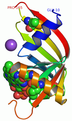
Chains, Units
Summary Information (see also Sequences/Alignments below) |
Ligands, Modified Residues, Ions (3, 6)| Asymmetric Unit (3, 6) Biological Unit 1 (2, 10) |
Sites (2, 2)
Asymmetric Unit (2, 2)
|
SS Bonds (0, 0)| (no "SS Bond" information available for 2HTI) |
Cis Peptide Bonds (0, 0)| (no "Cis Peptide Bond" information available for 2HTI) |
SAPs(SNPs)/Variants (0, 0)| (no "SAP(SNP)/Variant" information available for 2HTI) |
PROSITE Motifs (0, 0)| (no "PROSITE Motif" information available for 2HTI) |
Exons (0, 0)| (no "Exon" information available for 2HTI) |
Sequences/Alignments
Asymmetric UnitChain A from PDB Type:PROTEIN Length:126 aligned with Q9KFA8_BACHD | Q9KFA8 from UniProtKB/TrEMBL Length:184 Alignment length:156 19 29 39 49 59 69 79 89 99 109 119 129 139 149 159 Q9KFA8_BACHD 10 ECKDEKKITEFLNKARTGFLGLSTNDQPYVIPLNFVWHNHAIYFHGASEGRKIKMIEANPEVCFTICEDLGTIVSPVPAHTDTAYMSVIIFGTIEPVSAIEEGTEAMQQMLDKYVPGYYHSPLAASHVEKYRSSLGSRTAIYKISCRERTAKVNEP 165 SCOP domains d2htia1 A:10-165 Hypothetical protein BH0577 SCOP domains CATH domains 2htiA00 A:10-165 Electron Transport, Fmn-binding Protein; Chain A CATH domains Pfam domains ------------------------------------------------------------------------------------------------------------------------------------------------------------ Pfam domains SAPs(SNPs) ------------------------------------------------------------------------------------------------------------------------------------------------------------ SAPs(SNPs) PROSITE ------------------------------------------------------------------------------------------------------------------------------------------------------------ PROSITE Transcript ------------------------------------------------------------------------------------------------------------------------------------------------------------ Transcript 2hti A 10 ECKDEKKITEFLNKARTGFLGLSTNDQPYVIPLNFVWHNHAIYFHGASEGRKIKmIEANPEVCFTICEDL-------------AYmSVIIFGTIEPVSAIEEGTEAmQQmLDKYVP-----------------SLGSRTAIYKISCRERTAKVNEP 165 19 29 39 49 59 | 69 79 - | | 99 109 |119 | - - | 149 159 64-MSE 79 93 | 116-MSE 125 143 95-MSE 119-MSE
|
||||||||||||||||||||
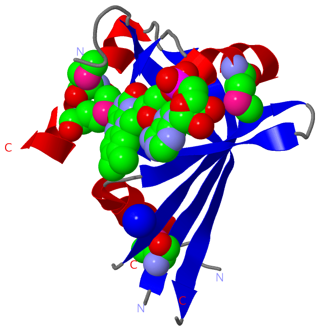
SCOP Domains (1, 1)
Asymmetric Unit

|
CATH Domains (1, 1)
Asymmetric Unit
|
Pfam Domains (0, 0)| (no "Pfam Domain" information available for 2HTI) |
Gene Ontology (4, 4)|
Asymmetric Unit(hide GO term definitions) Chain A (Q9KFA8_BACHD | Q9KFA8)
|
||||||||||||||||||||||||||||||||||||
Interactive Views
|
|||||||||||||||||||||||||||||||||||||||||||||||||||||||||||||||||||||||||||||||||||||||||||||||||||||||||||||||||||||||||||||||||||||||||||||||||||||||||||||
Still Images
|
||||||||||||||||
Databases
|
||||||||||||||||||||||||||||||||||||||||||||||||||||||||||||||||||||||||||||||||||||||||||||||||||||||||||||||||||||||||||||||||||||||||||||||||||||||||||||||||
Analysis Tools
|
|||||||||||||||||||||||||||||||||||||||||||||||||||||||||||||
Entries Sharing at Least One Protein Chain (UniProt ID)
Related Entries Specified in the PDB File
|
|