|
|
|
|
 Description
|
|
Compounds
|
||||||||||||||||||||||||||||||||||||||||||||||||
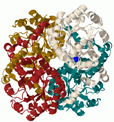
Chains, Units
Summary Information (see also Sequences/Alignments below) |
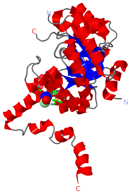
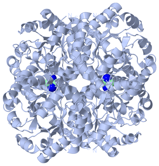
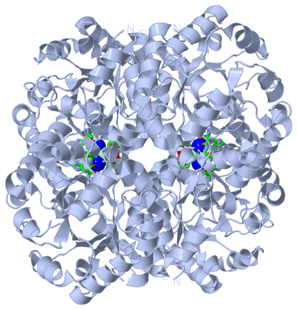
Ligands, Modified Residues, Ions (2, 3)| Asymmetric Unit (2, 3) Biological Unit 1 (0, 0) |
Sites (3, 3)
Asymmetric Unit (3, 3)
|
SS Bonds (0, 0)| (no "SS Bond" information available for 2HRW) |
Cis Peptide Bonds (1, 1)
Asymmetric Unit
|
||||||||
SAPs(SNPs)/Variants (0, 0)| (no "SAP(SNP)/Variant" information available for 2HRW) |
PROSITE Motifs (0, 0)| (no "PROSITE Motif" information available for 2HRW) |
Exons (0, 0)| (no "Exon" information available for 2HRW) |
Sequences/Alignments
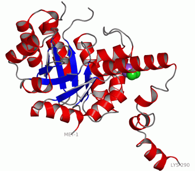
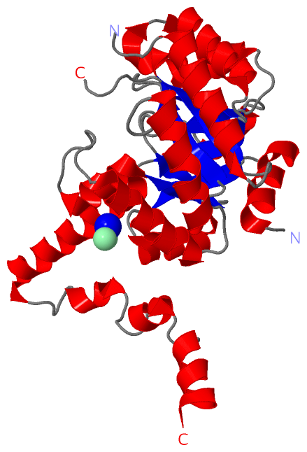
Asymmetric UnitChain A from PDB Type:PROTEIN Length:281 aligned with PPHA_VARSP | Q84G06 from UniProtKB/Swiss-Prot Length:290 Alignment length:290 10 20 30 40 50 60 70 80 90 100 110 120 130 140 150 160 170 180 190 200 210 220 230 240 250 260 270 280 290 PPHA_VARSP 1 MTKNQALRAALDSGRLFTAMAAHNPLVAKLAEQAGFGGIWGSGFELSASYAVPDANILSMSTHLEMMRAIASTVSIPLIADIDTGFGNAVNVHYVVPQYEAAGASAIVMEDKTFPKDTSLRTDGRQELVRIEEFQGKIAAATAARADRDFVVIARVEALIAGLGQQEAVRRGQAYEEAGADAILIHSRQKTPDEILAFVKSWPGKVPLVLVPTAYPQLTEADIAALSKVGIVIYGNHAIRAAVGAVREVFARIRRDGGIREVDAALPSVKEIIELQGDERMRAVEARYLK 290 SCOP domains d2hrwa_ A: automated matches SCOP domains CATH domains 2hrwA00 A:1-290 Phosphoenolpyruvate-binding domains CATH domains Pfam domains -------------------------------------------------------------------------------------------------------------------------------------------------------------------------------------------------------------------------------------------------------------------------------------------------- Pfam domains SAPs(SNPs) -------------------------------------------------------------------------------------------------------------------------------------------------------------------------------------------------------------------------------------------------------------------------------------------------- SAPs(SNPs) PROSITE -------------------------------------------------------------------------------------------------------------------------------------------------------------------------------------------------------------------------------------------------------------------------------------------------- PROSITE Transcript -------------------------------------------------------------------------------------------------------------------------------------------------------------------------------------------------------------------------------------------------------------------------------------------------- Transcript 2hrw A 1 MTKNQALRAALDSGRLFTAMAAHNPLVAKLAEQAGFGGIWGSGFELSASYAVPDANILSMSTHLEMMRAIASTVSIPLIADIDTGFGNAVNVHYVVPQYEAAGASAIVMEDKTFPK---------QELVRIEEFQGKIAAATAARADRDFVVIARVEALIAGLGQQEAVRRGQAYEEAGADAILIHSRQKTPDEILAFVKSWPGKVPLVLVPTAYPQLTEADIAALSKVGIVIYGNHAIRAAVGAVREVFARIRRDGGIREVDAALPSVKEIIELQGDERMRAVEARYLK 290 10 20 30 40 50 60 70 80 90 100 110 | - | 130 140 150 160 170 180 190 200 210 220 230 240 250 260 270 280 290 116 126
|
||||||||||||||||||||
SCOP Domains (1, 1)
Asymmetric Unit
 |
CATH Domains (1, 1)
Asymmetric Unit
|
Pfam Domains (0, 0)| (no "Pfam Domain" information available for 2HRW) |
Gene Ontology (4, 4)|
Asymmetric Unit(hide GO term definitions) Chain A (PPHA_VARSP | Q84G06)
|
||||||||||||||||||||||||||||||
Interactive Views
|
||||||||||||||||||||||||||||||||||||||||||||||||||||||||||||||||||||||||||||||||||||||||||||||||||||||||||||||||||||||||||||||||||||||||||||||||||||||||||||||
Still Images
|
||||||||||||||||
Databases
|
||||||||||||||||||||||||||||||||||||||||||||||||||||||||||||||||||||||||||||||||||||||||||||||||||||||||||||||||||||||||||||||||||||||||||||||||||||||||||||||||
Analysis Tools
|
|||||||||||||||||||||||||||||||||||||||||||||||||||||||||||||
Entries Sharing at Least One Protein Chain (UniProt ID)
Related Entries Specified in the PDB File
|
|