|
|
|
|
 Description
|
|
Compounds
|
||||||||||||||||||||||||||||||||||||||||
Chains, Units
Summary Information (see also Sequences/Alignments below) |
Ligands, Modified Residues, Ions (3, 7)| Asymmetric Unit (3, 7) Biological Unit 1 (1, 6) |
Sites (3, 3)
Asymmetric Unit (3, 3)
|
SS Bonds (0, 0)| (no "SS Bond" information available for 2HKV) |
Cis Peptide Bonds (0, 0)| (no "Cis Peptide Bond" information available for 2HKV) |
SAPs(SNPs)/Variants (0, 0)| (no "SAP(SNP)/Variant" information available for 2HKV) |
PROSITE Motifs (0, 0)| (no "PROSITE Motif" information available for 2HKV) |
Exons (0, 0)| (no "Exon" information available for 2HKV) |
Sequences/Alignments
Asymmetric UnitChain A from PDB Type:PROTEIN Length:148 aligned with B1YEV7_EXIS2 | B1YEV7 from UniProtKB/TrEMBL Length:148 Alignment length:148 1 | 9 19 29 39 49 59 69 79 89 99 109 119 129 139 B1YEV7_EXIS2 - -MTDWQQALDRHVGVGVRTTRDLIRLIQPEDWDKRPISGKRSVYEVAVHLAVLLEADLRIATGATADEMAQFYAVPVLPEQLVDRLDQSWQYYQDRLMADFSTETTYWGVTDSTTGWLLEAAVHLYHHRSQLLDYLNLLGYDIKLDLF 147 SCOP domains -d2hkva1 A:1-147 Hypothetical protein ExigDRAFT_2445 SCOP domains CATH domains 2hkvA01 A:0-140 dinb family like domain ------- CATH domains Pfam domains ---------------------------------------------------------------------------------------------------------------------------------------------------- Pfam domains SAPs(SNPs) ---------------------------------------------------------------------------------------------------------------------------------------------------- SAPs(SNPs) PROSITE ---------------------------------------------------------------------------------------------------------------------------------------------------- PROSITE Transcript ---------------------------------------------------------------------------------------------------------------------------------------------------- Transcript 2hkv A 0 GmTDWQQALDRHVGVGVRTTRDLIRLIQPEDWDKRPISGKRSVYEVAVHLAVLLEADLRIATGATADEmAQFYAVPVLPEQLVDRLDQSWQYYQDRLmADFSTETTYWGVTDSTTGWLLEAAVHLYHHRSQLLDYLNLLGYDIKLDLF 147 | 9 19 29 39 49 59 69 79 89 |99 109 119 129 139 | 68-MSE 97-MSE 1-MSE
|
||||||||||||||||||||
SCOP Domains (1, 1)
Asymmetric Unit
 |
CATH Domains (1, 1)
Asymmetric Unit
|
Pfam Domains (0, 0)| (no "Pfam Domain" information available for 2HKV) |
Gene Ontology (0, 0)|
Asymmetric Unit(hide GO term definitions)
(no "Gene Ontology" information available for 2HKV)
|
Interactive Views
|
||||||||||||||||||||||||||||||||||||||||||||||||||||||||||||||||||||||||||||||||||||||||||||||||||||||||||||||||||||||||||||||||||||||||||||||||||||||||||||||||||||
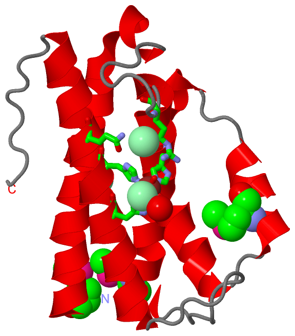
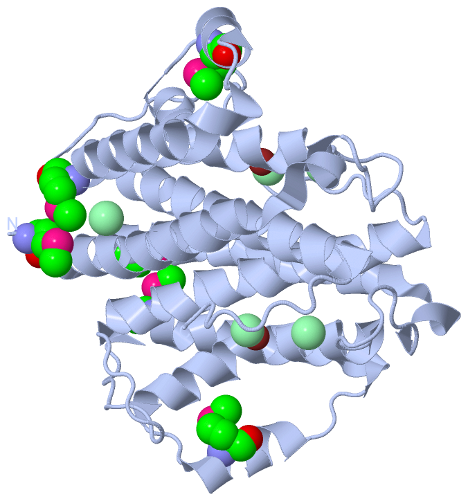
Still Images
|
||||||||||||||||
Databases
|
||||||||||||||||||||||||||||||||||||||||||||||||||||||||||||||||||||||||||||||||||||||||||||||||||||||||||||||||||||||||||||||||||||||||||||||||||||||||||||||||
Analysis Tools
|
|||||||||||||||||||||||||||||||||||||||||||||||||||||||||||||
Entries Sharing at Least One Protein Chain (UniProt ID)
Related Entries Specified in the PDB File
|
|