Show PDB file:   
         Plain Text   HTML   (compressed file size)
QuickSearch:   
by PDB,NDB,UniProt,PROSITE Code or Search Term(s)  
(-)Asymmetric Unit
(-)Asym. Unit - sites
(-)Biological Unit 1
(-)Biol. Unit 1 - sites
(-)Biological Unit 2
collapse expand < >
Image Asymmetric Unit
Asymmetric Unit  (Jmol Viewer)
Image Asym. Unit - sites
Asym. Unit - sites  (Jmol Viewer)
Image Biological Unit 1
Biological Unit 1  (Jmol Viewer)
Image Biol. Unit 1 - sites
Biol. Unit 1 - sites  (Jmol Viewer)
Image Biological Unit 2
Biological Unit 2  (Jmol Viewer)

(-) Description

Title :  CRYSTAL STRUCTURE OF A PYRIDOXAMINE 5'-PHOSPHATE OXIDASE-RELATED PROTEIN (SSUIDRAFT_2804) FROM STREPTOCOCCUS SUIS 89/1591 AT 2.00 A RESOLUTION
 
Authors :  Joint Center For Structural Genomics (Jcsg)
Date :  28 Jun 06  (Deposition) - 15 Aug 06  (Release) - 13 Jul 11  (Revision)
Method :  X-RAY DIFFRACTION
Resolution :  2.00
Chains :  Asym. Unit :  A
Biol. Unit 1:  A  (2x)
Biol. Unit 2:  A  (1x)
Keywords :  Structural Genomics, Joint Center For Structural Genomics, Jcsg, Protein Structure Initiative, Psi-2, Oxidoreductase (Keyword Search: [Gene Ontology, PubMed, Web (Google))
 
Reference :  Joint Center For Structural Genomics (Jcsg)
Crystal Structure Of Pyridoxamine 5'-Phosphate Oxidase-Related (Zp_00875725. 1) From Streptococcus Suis 89-1591 At 2. 00 A Resolution
To Be Published
PubMed: search

(-) Compounds

Molecule 1 - PYRIDOXAMINE 5'-PHOSPHATE OXIDASE-RELATED
    ChainsA
    EC Number1.4.3.5
    EngineeredYES
    Expression SystemESCHERICHIA COLI
    Expression System Taxid562
    Expression System Vector TypePLASMID
    GeneZP_00875725.1
    Organism ScientificSTREPTOCOCCUS SUIS
    Organism Taxid286604
    Strain89/1591

 Structural Features

(-) Chains, Units

  1
Asymmetric Unit A
Biological Unit 1 (2x)A
Biological Unit 2 (1x)A

Summary Information (see also Sequences/Alignments below)

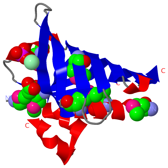
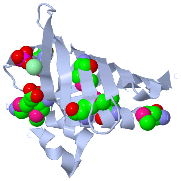
(-) Ligands, Modified Residues, Ions  (4, 10)

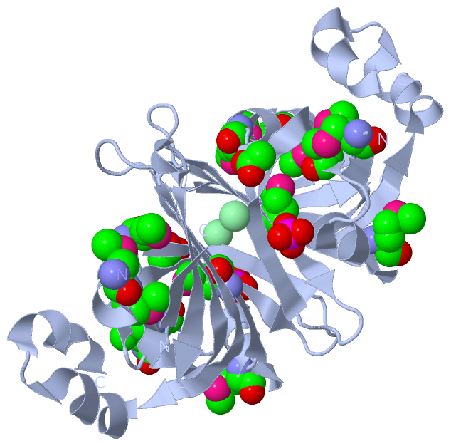
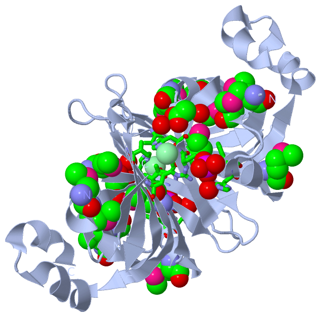
Asymmetric Unit (4, 10)
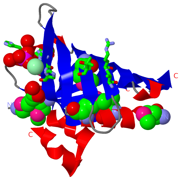
No.NameCountTypeFull Name
1ACT1Ligand/IonACETATE ION
2CL1Ligand/IonCHLORIDE ION
3MSE7Mod. Amino AcidSELENOMETHIONINE
4PO41Ligand/IonPHOSPHATE ION
Biological Unit 1 (3, 18)
No.NameCountTypeFull Name
1ACT2Ligand/IonACETATE ION
2CL-1Ligand/IonCHLORIDE ION
3MSE14Mod. Amino AcidSELENOMETHIONINE
4PO42Ligand/IonPHOSPHATE ION
Biological Unit 2 (3, 9)
No.NameCountTypeFull Name
1ACT1Ligand/IonACETATE ION
2CL-1Ligand/IonCHLORIDE ION
3MSE7Mod. Amino AcidSELENOMETHIONINE
4PO41Ligand/IonPHOSPHATE ION

(-) Sites  (3, 3)

Asymmetric Unit (3, 3)
No.NameEvidenceResiduesDescription
1AC1SOFTWAREARG A:30 , HIS A:51 , PHE A:52 , ARG A:78 , HOH A:170 , HOH A:188BINDING SITE FOR RESIDUE PO4 A 150
2AC2SOFTWAREHIS A:31 , THR A:65 , VAL A:76 , ARG A:78BINDING SITE FOR RESIDUE CL A 151
3AC3SOFTWAREHIS A:31 , HOH A:195 , HOH A:197BINDING SITE FOR RESIDUE ACT A 152

(-) SS Bonds  (0, 0)

(no "SS Bond" information available for 2HHZ)

(-) Cis Peptide Bonds  (0, 0)

(no "Cis Peptide Bond" information available for 2HHZ)

 Sequence-Structure Mapping

(-) SAPs(SNPs)/Variants  (0, 0)

(no "SAP(SNP)/Variant" information available for 2HHZ)

(-) PROSITE Motifs  (0, 0)

(no "PROSITE Motif" information available for 2HHZ)

(-) Exons   (0, 0)

(no "Exon" information available for 2HHZ)

(-) Sequences/Alignments

Asymmetric Unit
   Reformat: Number of residues per line =  ('0' or empty: single-line sequence representation)
  Number of residues per labelling interval =   
  UniProt sequence: complete  aligned part    
   Show mapping: SCOP domains CATH domains Pfam domains Secondary structure (by author)
SAPs(SNPs) PROSITE motifs Exons
(details for a mapped element are shown in a popup box when the mouse pointer rests over it)
Chain A from PDB  Type:PROTEIN  Length:139
                                                                                                                                                                           
               SCOP domains ------------------------------------------------------------------------------------------------------------------------------------------- SCOP domains
               CATH domains -2hhzA00 A:2-149 Electron Transport, Fmn-binding Protein; Chain A                                                                           CATH domains
               Pfam domains ------------------------------------------------------------------------------------------------------------------------------------------- Pfam domains
         Sec.struct. author .hhhhhhhhhhhh.eeeeeee.....eeeeeeeeeeee..eeeeee...hhhhhhhhhh.eeeeeeee......eeeeeeeeeeeehhhhhhhhhh.hhhhhhhh...eeeeeeeeeeeeee.hhheeeeeee..eee. Sec.struct. author
                 SAPs(SNPs) ------------------------------------------------------------------------------------------------------------------------------------------- SAPs(SNPs)
                    PROSITE ------------------------------------------------------------------------------------------------------------------------------------------- PROSITE
                 Transcript ------------------------------------------------------------------------------------------------------------------------------------------- Transcript
                 2hhz A   1 mELKDImHILEDmKVGVFATLDEYGNPHARHAHITAANEEGIFFmTSPETHFYDQLmGDQRVAmTAISEEGYLIQVVRVEGTARPVENDYLKTVFADNPYYQHIYKTmQVFQIYAGHGFYHSLTQGHKYIFSIGEVRAL 149
                            |     | 10  |     20        30        40    |   50      | 60   |    70        80        90       100     ||115       125       135   ||    
                            |     7-MSE |                              45-MSE      57-MSE 64-MSE                                   106||                       139|    
                            1-MSE      13-MSE                                                                                       112|                        145    
                                                                                                                                     113-MSE                           

   Legend:   → Mismatch (orange background)
  - → Gap (green background, '-', border residues have a numbering label)
    → Modified Residue (blue background, lower-case, 'x' indicates undefined single-letter code, labelled with number + name)
  x → Chemical Group (purple background, 'x', labelled with number + name, e.g. ACE or NH2)
  extra numbering lines below/above indicate numbering irregularities and modified residue names etc., number ends below/above '|'

 Classification and Annotation

(-) SCOP Domains  (0, 0)

(no "SCOP Domain" information available for 2HHZ)

(-) CATH Domains  (1, 1)

Asymmetric Unit
(-)
Class: Mainly Beta (13760)

(-) Pfam Domains  (0, 0)

(no "Pfam Domain" information available for 2HHZ)

(-) Gene Ontology  (0, 0)

Asymmetric Unit(hide GO term definitions)
    (no "Gene Ontology" information available for 2HHZ)

 Visualization

(-) Interactive Views

Asymmetric Unit
  Complete Structure
    Jena3D(integrated viewing of ligand, site, SAP, PROSITE, SCOP information)
    WebMol | AstexViewer[tm]@PDBe
(Java Applets, require no local installation except for Java; loading may be slow)
    STRAP
(Java WebStart application, automatic local installation, requires Java; full application with system access!)
    RasMol
(require local installation)
    Molscript (VRML)
(requires installation of a VRML viewer; select preferred view via VRML and generate a mono or stereo PDF format file)
 
  Ligands, Modified Residues, Ions
    ACT  [ RasMol | Jena3D ]  +environment [ RasMol | Jena3D ]
    CL  [ RasMol | Jena3D ]  +environment [ RasMol | Jena3D ]
    MSE  [ RasMol | Jena3D ]  +environment [ RasMol | Jena3D ]
    PO4  [ RasMol | Jena3D ]  +environment [ RasMol | Jena3D ]
 
  Sites
    AC1  [ RasMol ]  +environment [ RasMol ]
    AC2  [ RasMol ]  +environment [ RasMol ]
    AC3  [ RasMol ]  +environment [ RasMol ]
 
  Cis Peptide Bonds
(no "Cis Peptide Bonds" information available for 2hhz)
 
Biological Units
  Complete Structure
    Biological Unit 1  [ Jena3D ]
    Biological Unit 2  [ Jena3D ]

(-) Still Images

Jmol
  protein: cartoon or spacefill or dots and stick; nucleic acid: cartoon and stick; ligands: spacefill; active site: stick
Molscript
  protein, nucleic acid: cartoon; ligands: spacefill; active site: ball and stick

 Databases and Analysis Tools

(-) Databases

Access by PDB/NDB ID
  2hhz
    Family and Domain InformationProDom | SYSTERS
    General Structural InformationGlycoscienceDB | MMDB | NDB | OCA | PDB | PDBe | PDBj | PDBsum | PDBWiki | PQS | PROTEOPEDIA
    Orientation in MembranesOPM
    Protein SurfaceSURFACE
    Secondary StructureDSSP (structure derived) | HSSP (homology derived)
    Structural GenomicsGeneCensus
    Structural NeighboursCE | VAST
    Structure ClassificationCATH | Dali | SCOP
    Validation and Original DataBMRB Data View | BMRB Restraints Grid | EDS | PROCHECK | RECOORD | WHAT_CHECK
 
Access by UniProt ID/Accession number
  (no 'UniProt ID/Accession number' available) |
    Comparative Protein Structure ModelsModBase
    Genomic InformationEnsembl
    Protein-protein InteractionDIP
    Sequence, Family and Domain InformationInterPro | Pfam | SMART | UniProt
 
Access by Enzyme Classificator   (EC Number)
  1.4.3.5
    General Enzyme InformationBRENDA | EC-PDB | Enzyme | IntEnz
    PathwayKEGG | MetaCyc
 
Access by Disease Identifier   (MIM ID)
  (no 'MIM ID' available)
    Disease InformationOMIM
 
Access by GenAge ID
  (no 'GenAge ID' available)
    Age Related InformationGenAge

(-) Analysis Tools

Access by PDB/NDB ID
    Domain InformationXDom
    Interatomic Contacts of Structural UnitsCSU
    Ligand-protein ContactsLPC
    Protein CavitiescastP
    Sequence and Secondary StructurePDBCartoon
    Structure AlignmentSTRAP(Java WebStart application, automatic local installation, requires Java; full application with system access!)
    Structure and Sequence BrowserSTING
 
Access by UniProt ID/Accession number
  (no 'UniProt ID/Accession number' available) |
    Protein Disorder PredictionDisEMBL | FoldIndex | GLOBPLOT

 Related Entries

(-) Entries Sharing at Least One Protein Chain (UniProt ID)

(no "Entries Sharing at Least One Protein Chain" available for 2HHZ)

(-) Related Entries Specified in the PDB File

(no "Related Entries Specified in the PDB File" available for 2HHZ)