|
|
|
|
 Description
|
|
Compounds
|
||||||||||||||||||||||||||||||||||||||||||||||||
Chains, Units
Summary Information (see also Sequences/Alignments below) |
Ligands, Modified Residues, Ions (0, 0)| (no "Ligand,Modified Residues,Ions" information available for 2HH8) |
Sites (0, 0)| (no "Site" information available for 2HH8) |
SS Bonds (0, 0)| (no "SS Bond" information available for 2HH8) |
Cis Peptide Bonds (0, 0)| (no "Cis Peptide Bond" information available for 2HH8) |
SAPs(SNPs)/Variants (0, 0)| (no "SAP(SNP)/Variant" information available for 2HH8) |
PROSITE Motifs (0, 0)| (no "PROSITE Motif" information available for 2HH8) |
Exons (0, 0)| (no "Exon" information available for 2HH8) |
Sequences/Alignments
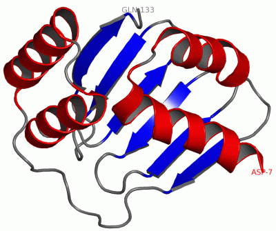
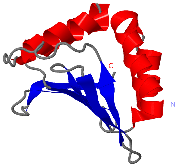
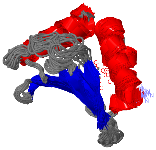
NMR StructureChain A from PDB Type:PROTEIN Length:127 aligned with YDFO_ECOLI | P76156 from UniProtKB/Swiss-Prot Length:136 Alignment length:127 11 21 31 41 51 61 71 81 91 101 111 121 YDFO_ECOLI 2 DQVVIFKQIFDKVRNDLNYQWFYSELKRHNVSHYIYYLATENVHIVLKNDNTVLLKGLKNIVSVKFSKDRHLIETTSNKLKSREITFQEYRRNLAKAGVFRWVTNIHEQKRYYYTFDNSLLFTESIQ 128 SCOP domains d2hh8a1 A:7-133 Hypothetical protein YdfO SCOP domains CATH domains 2hh8A00 A:7-133 YdfO-like domain CATH domains Pfam domains ------------------------------------------------------------------------------------------------------------------------------- Pfam domains SAPs(SNPs) ------------------------------------------------------------------------------------------------------------------------------- SAPs(SNPs) PROSITE ------------------------------------------------------------------------------------------------------------------------------- PROSITE Transcript ------------------------------------------------------------------------------------------------------------------------------- Transcript 2hh8 A 7 DQVVIFKQIFDKVRNDLNYQWFYSELKRHNVSHYIYYLATENVHIVLKNDNTVLLKGLKNIVSVKFSKDRHLIETTSNKLKSREITFQEYRRNLAKAGVFRWVTNIHEQKRYYYTFDNSLLFTESIQ 133 16 26 36 46 56 66 76 86 96 106 116 126
|
||||||||||||||||||||
SCOP Domains (1, 1)
NMR Structure

|
CATH Domains (1, 1)
NMR Structure
|
Pfam Domains (0, 0)| (no "Pfam Domain" information available for 2HH8) |
Gene Ontology (0, 0)|
NMR Structure(hide GO term definitions)
(no "Gene Ontology" information available for 2HH8)
|
Interactive Views
|
||||||||||||||||||||||||||||||||||||||||||||||||||||||||||||||||||||||||||||||||||||||||||||||||||||||||||||||||||||
Still Images
|
||||||||||||||||
Databases
|
||||||||||||||||||||||||||||||||||||||||||||||||||||||||||||||||||||||||||||||||||||||||||||||||||||||||||||||||||||||||||||||||||||||||||||||||||||||||||||||||
Analysis Tools
|
|||||||||||||||||||||||||||||||||||||||||||||||||||||||||||||
Entries Sharing at Least One Protein Chain (UniProt ID)
Related Entries Specified in the PDB File
|
|