|
|
|
|
 Description
|
|
Compounds
|
||||||||||||||||||||||||||||||||||||
Chains, Units
Summary Information (see also Sequences/Alignments below) |
Ligands, Modified Residues, Ions (1, 6)
Asymmetric Unit (1, 6)
|
Sites (0, 0)| (no "Site" information available for 2HH6) |
SS Bonds (0, 0)| (no "SS Bond" information available for 2HH6) |
Cis Peptide Bonds (0, 0)| (no "Cis Peptide Bond" information available for 2HH6) |
SAPs(SNPs)/Variants (0, 0)| (no "SAP(SNP)/Variant" information available for 2HH6) |
PROSITE Motifs (0, 0)| (no "PROSITE Motif" information available for 2HH6) |
Exons (0, 0)| (no "Exon" information available for 2HH6) |
Sequences/Alignments
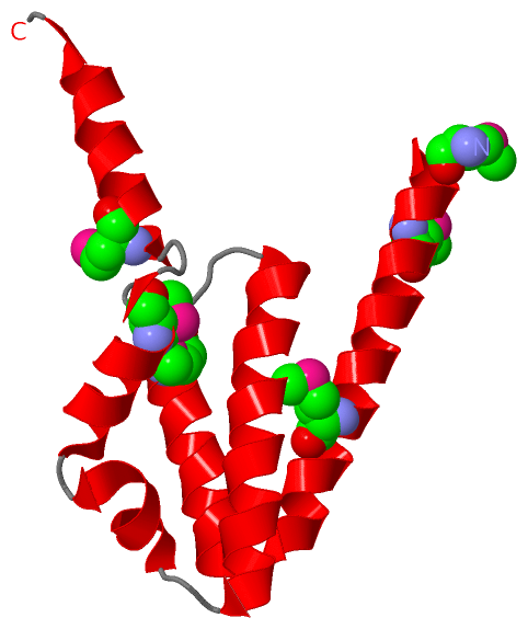
Asymmetric UnitChain A from PDB Type:PROTEIN Length:112 aligned with Q9K5V7_BACHD | Q9K5V7 from UniProtKB/TrEMBL Length:112 Alignment length:112 10 20 30 40 50 60 70 80 90 100 110 Q9K5V7_BACHD 1 MSFIEKMIGSLNDKREWKAMEARAKALPKEYHHAYKAIQKYMWTSGGPTDWQDTKRIFGGILDLFEEGAAEGKKVTDLTGEDVAAFCDELMKDTKTWMDKYRTKLNDSIGRD 112 SCOP domains d2hh6a1 A:1-112 Uncharacterized protein BH3980 SCOP domains CATH domains -2hh6A00 A:2-112 c-terminal domain of poly(a) binding protein CATH domains Pfam domains ---------------------------------------------------------------------------------------------------------------- Pfam domains SAPs(SNPs) ---------------------------------------------------------------------------------------------------------------- SAPs(SNPs) PROSITE ---------------------------------------------------------------------------------------------------------------- PROSITE Transcript ---------------------------------------------------------------------------------------------------------------- Transcript 2hh6 A 1 mSFIEKmIGSLNDKREWKAmEARAKALPKEYHHAYKAIQKYmWTSGGPTDWQDTKRIFGGILDLFEEGAAEGKKVTDLTGEDVAAFCDELmKDTKTWmDKYRTKLNDSIGRD 112 | | 10 20 30 40 | 50 60 70 80 90| 100 110 | 7-MSE 20-MSE 42-MSE 91-MSE 98-MSE 1-MSE
|
||||||||||||||||||||
SCOP Domains (1, 1)
Asymmetric Unit
 |
CATH Domains (1, 1)
Asymmetric Unit
|
Pfam Domains (0, 0)| (no "Pfam Domain" information available for 2HH6) |
Gene Ontology (1, 1)|
Asymmetric Unit(hide GO term definitions) Chain A (Q9K5V7_BACHD | Q9K5V7)
|
||||||||||||
Interactive Views
|
|||||||||||||||||||||||||||||||||||||||||||||||||||||||||||||||||||||||||||||||||||||||||||||||||||||||||||||||||||||||||||||||||||||||
Still Images
|
||||||||||||||||
Databases
|
||||||||||||||||||||||||||||||||||||||||||||||||||||||||||||||||||||||||||||||||||||||||||||||||||||||||||||||||||||||||||||||||||||||||||||||||||||||||||||||||
Analysis Tools
|
|||||||||||||||||||||||||||||||||||||||||||||||||||||||||||||
Entries Sharing at Least One Protein Chain (UniProt ID)
Related Entries Specified in the PDB File
|
|