|
|
|
|
 Description
|
|
Compounds
|
||||||||||||||||||||||||||||||||||||||||||||||||||||||||
Chains, Units
Summary Information (see also Sequences/Alignments below) |
Ligands, Modified Residues, Ions (1, 1)
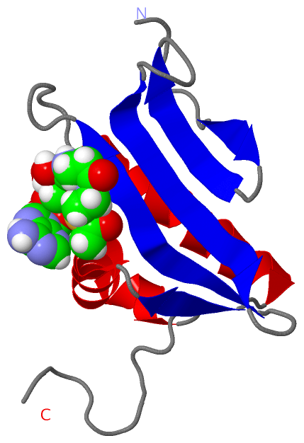
NMR Structure (1, 1)
|
Sites (1, 1)
NMR Structure (1, 1)
|
SS Bonds (0, 0)| (no "SS Bond" information available for 2H5M) |
Cis Peptide Bonds (0, 0)| (no "Cis Peptide Bond" information available for 2H5M) |
SAPs(SNPs)/Variants (0, 0)| (no "SAP(SNP)/Variant" information available for 2H5M) |
PROSITE Motifs (0, 0)| (no "PROSITE Motif" information available for 2H5M) |
Exons (0, 0)| (no "Exon" information available for 2H5M) |
Sequences/Alignments
NMR StructureChain A from PDB Type:PROTEIN Length:102 aligned with A0A0H2WXG3_S | A0A0H2WXG3 from UniProtKB/TrEMBL Length:94 Alignment length:102 94 10 20 30 40 50 60 70 80 90 | - A0A0H2WXG3_S 1 MSNLEIKQGENKFYIGDDENNALAEITYRFVDNNEINIDHTGVSDELGGQGVGKKLLKAVVEHARENNLKIIASCSFAKHMLEKEDSYQDVYLG-------- - SCOP domains d2h5ma_ A: Hypothetical protein SA2309 SCOP domains CATH domains 2h5mA00 A:1-102 [code=3.40.630.30, no name defined] CATH domains Pfam domains ------------------------------------------------------------------------------------------------------ Pfam domains SAPs(SNPs) ------------------------------------------------------------------------------------------------------ SAPs(SNPs) PROSITE ------------------------------------------------------------------------------------------------------ PROSITE Transcript ------------------------------------------------------------------------------------------------------ Transcript 2h5m A 1 MSNLEIKQGENKFYIGDDENNALAEITYRFVDNNEINIDHTGVSDELGGQGVGKKLLKAVVEHARENNLKIIASCSFAKHMLEKEDSYQDVYLGLEHHHHHH 102 10 20 30 40 50 60 70 80 90 100
|
||||||||||||||||||||
SCOP Domains (1, 1)
NMR Structure
 |
CATH Domains (1, 1)
NMR Structure
|
Pfam Domains (0, 0)| (no "Pfam Domain" information available for 2H5M) |
Gene Ontology (0, 0)|
NMR Structure(hide GO term definitions)
(no "Gene Ontology" information available for 2H5M)
|
Interactive Views
|
||||||||||||||||||||||||||||||||||||||||||||||||||||||||||||||||||||||||||||||||||||||||||||||||||||||||||||||||||||||
Still Images
|
||||||||||||||||
Databases
|
||||||||||||||||||||||||||||||||||||||||||||||||||||||||||||||||||||||||||||||||||||||||||||||||||||||||||||||||||||||||||||||||||||||||||||||||||||||||||||||||
Analysis Tools
|
|||||||||||||||||||||||||||||||||||||||||||||||||||||||||||||
Entries Sharing at Least One Protein Chain (UniProt ID)
Related Entries Specified in the PDB File
|
|