|
|
|
|
 Description
|
|
Compounds
|
||||||||||||||||||||||||||||||||||||||||||||||||||||||||
Chains, Units
Summary Information (see also Sequences/Alignments below) |
Ligands, Modified Residues, Ions (0, 0)| (no "Ligand,Modified Residues,Ions" information available for 2H2Y) |
Sites (0, 0)| (no "Site" information available for 2H2Y) |
SS Bonds (1, 1)
Asymmetric Unit
|
||||||||
Cis Peptide Bonds (4, 4)
Asymmetric Unit
|
||||||||||||||||||||
SAPs(SNPs)/Variants (0, 0)| (no "SAP(SNP)/Variant" information available for 2H2Y) |
PROSITE Motifs (0, 0)| (no "PROSITE Motif" information available for 2H2Y) |
Exons (0, 0)| (no "Exon" information available for 2H2Y) |
Sequences/Alignments
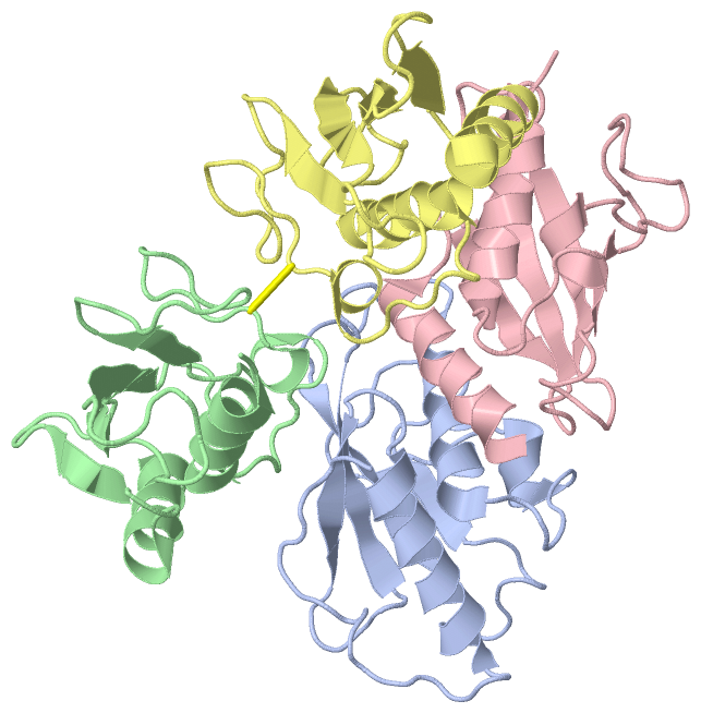
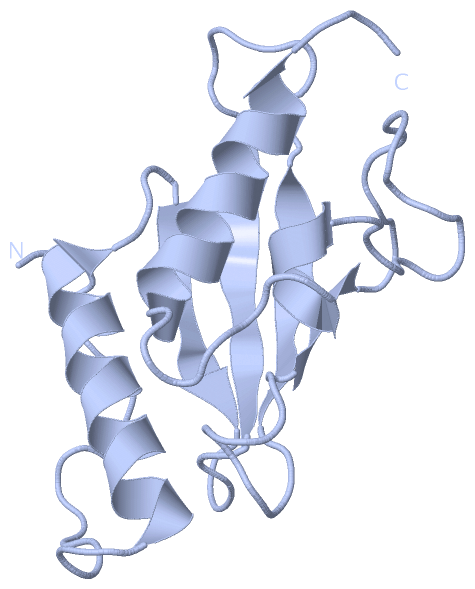
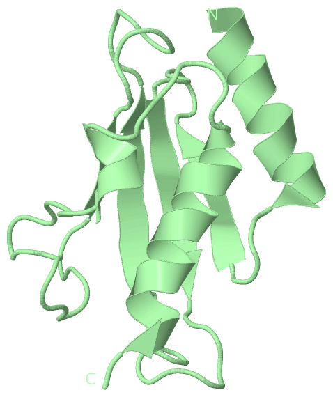
Asymmetric UnitChain A from PDB Type:PROTEIN Length:133 aligned with Q8IDP1_PLAF7 | Q8IDP1 from UniProtKB/TrEMBL Length:278 Alignment length:133 126 136 146 156 166 176 186 196 206 216 226 236 246 Q8IDP1_PLAF7 117 KPSRTVEKHIKTKYNLGNANYRIQKELNNFLKNPPINCTIDVHPSNIRIWIVQYVGLENTIYANEVYKIKIIFPDNYPLKPPIVYFLQKPPKHTHVYSNGDICLSVLGDDYNPSLSISGLILSIISMLSSAKE 249 SCOP domains ------------------------------------------------------------------------------------------------------------------------------------- SCOP domains CATH domains ------------------------------------------------------------------------------------------------------------------------------------- CATH domains Pfam domains ------------------------------------------------------------------------------------------------------------------------------------- Pfam domains SAPs(SNPs) ------------------------------------------------------------------------------------------------------------------------------------- SAPs(SNPs) PROSITE ------------------------------------------------------------------------------------------------------------------------------------- PROSITE Transcript ------------------------------------------------------------------------------------------------------------------------------------- Transcript 2h2y A 3 KPSRTVEKHIKTKYNLGNANYRIQKELNNFLKNPPINCTIDVHPSNIRIWIVQYVGLENTIYANEVYKIKIIFPDNYPLKPPIVYFLQKPPKHTHVYSNGDICLSVLGDDYNPSLSISGLILSIISMLSSAKE 135 12 22 32 42 52 62 72 82 92 102 112 122 132 Chain B from PDB Type:PROTEIN Length:113 aligned with Q8IDP1_PLAF7 | Q8IDP1 from UniProtKB/TrEMBL Length:278 Alignment length:113 145 155 165 175 185 195 205 215 225 235 245 Q8IDP1_PLAF7 136 NYRIQKELNNFLKNPPINCTIDVHPSNIRIWIVQYVGLENTIYANEVYKIKIIFPDNYPLKPPIVYFLQKPPKHTHVYSNGDICLSVLGDDYNPSLSISGLILSIISMLSSAK 248 SCOP domains ----------------------------------------------------------------------------------------------------------------- SCOP domains CATH domains ----------------------------------------------------------------------------------------------------------------- CATH domains Pfam domains ----------------------------------------------------------------------------------------------------------------- Pfam domains SAPs(SNPs) ----------------------------------------------------------------------------------------------------------------- SAPs(SNPs) PROSITE ----------------------------------------------------------------------------------------------------------------- PROSITE Transcript ----------------------------------------------------------------------------------------------------------------- Transcript 2h2y B 22 NYRIQKELNNFLKNPPINCTIDVHPSNIRIWIVQYVGLENTIYANEVYKIKIIFPDNYPLKPPIVYFLQKPPKHTHVYSNGDICLSVLGDDYNPSLSISGLILSIISMLSSAK 134 31 41 51 61 71 81 91 101 111 121 131 Chain C from PDB Type:PROTEIN Length:113 aligned with Q8IDP1_PLAF7 | Q8IDP1 from UniProtKB/TrEMBL Length:278 Alignment length:113 145 155 165 175 185 195 205 215 225 235 245 Q8IDP1_PLAF7 136 NYRIQKELNNFLKNPPINCTIDVHPSNIRIWIVQYVGLENTIYANEVYKIKIIFPDNYPLKPPIVYFLQKPPKHTHVYSNGDICLSVLGDDYNPSLSISGLILSIISMLSSAK 248 SCOP domains ----------------------------------------------------------------------------------------------------------------- SCOP domains CATH domains ----------------------------------------------------------------------------------------------------------------- CATH domains Pfam domains ----------------------------------------------------------------------------------------------------------------- Pfam domains SAPs(SNPs) ----------------------------------------------------------------------------------------------------------------- SAPs(SNPs) PROSITE ----------------------------------------------------------------------------------------------------------------- PROSITE Transcript ----------------------------------------------------------------------------------------------------------------- Transcript 2h2y C 22 NYRIQKELNNFLKNPPINCTIDVHPSNIRIWIVQYVGLENTIYANEVYKIKIIFPDNYPLKPPIVYFLQKPPKHTHVYSNGDICLSVLGDDYNPSLSISGLILSIISMLSSAK 134 31 41 51 61 71 81 91 101 111 121 131 Chain D from PDB Type:PROTEIN Length:112 aligned with Q8IDP1_PLAF7 | Q8IDP1 from UniProtKB/TrEMBL Length:278 Alignment length:112 145 155 165 175 185 195 205 215 225 235 245 Q8IDP1_PLAF7 136 NYRIQKELNNFLKNPPINCTIDVHPSNIRIWIVQYVGLENTIYANEVYKIKIIFPDNYPLKPPIVYFLQKPPKHTHVYSNGDICLSVLGDDYNPSLSISGLILSIISMLSSA 247 SCOP domains ---------------------------------------------------------------------------------------------------------------- SCOP domains CATH domains ---------------------------------------------------------------------------------------------------------------- CATH domains Pfam domains ---------------------------------------------------------------------------------------------------------------- Pfam domains SAPs(SNPs) ---------------------------------------------------------------------------------------------------------------- SAPs(SNPs) PROSITE ---------------------------------------------------------------------------------------------------------------- PROSITE Transcript ---------------------------------------------------------------------------------------------------------------- Transcript 2h2y D 22 NYRIQKELNNFLKNPPINCTIDVHPSNIRIWIVQYVGLENTIYANEVYKIKIIFPDNYPLKPPIVYFLQKPPKHTHVYSNGDICLSVLGDDYNPSLSISGLILSIISMLSSA 133 31 41 51 61 71 81 91 101 111 121 131
|
||||||||||||||||||||
SCOP Domains (0, 0)| (no "SCOP Domain" information available for 2H2Y) |
CATH Domains (0, 0)| (no "CATH Domain" information available for 2H2Y) |
Pfam Domains (0, 0)| (no "Pfam Domain" information available for 2H2Y) |
Gene Ontology (12, 12)|
Asymmetric Unit(hide GO term definitions) Chain A,B,C,D (Q8IDP1_PLAF7 | Q8IDP1)
|
||||||||||||||||||||||||||||||||||||||||||||||||||||||||||||||||||||||||||||||||||||||||||
Interactive Views
|
|||||||||||||||||||||||||||||||||||||||||||||||||||||||||||||||||||||||||||||||||||||||||||||||||||||||||||||||||||||||||||||||||||||||||||||||||||||||||||||||||||||||||||
Still Images
|
||||||||||||||||
Databases
|
||||||||||||||||||||||||||||||||||||||||||||||||||||||||||||||||||||||||||||||||||||||||||||||||||||||||||||||||||||||||||||||||||||||||||||||||||||||||||||||||
Analysis Tools
|
|||||||||||||||||||||||||||||||||||||||||||||||||||||||||||||
Entries Sharing at Least One Protein Chain (UniProt ID)
Related Entries Specified in the PDB File
|
|