|
|
|
|
 Description
|
|
Compounds
|
||||||||||||||||||||||||||||||||||||||||||||||||||||
Chains, Units
Summary Information (see also Sequences/Alignments below) |
Ligands, Modified Residues, Ions (0, 0)| (no "Ligand,Modified Residues,Ions" information available for 2GOV) |
Sites (0, 0)| (no "Site" information available for 2GOV) |
SS Bonds (0, 0)| (no "SS Bond" information available for 2GOV) |
Cis Peptide Bonds (0, 0)| (no "Cis Peptide Bond" information available for 2GOV) |
SAPs(SNPs)/Variants (0, 0)| (no "SAP(SNP)/Variant" information available for 2GOV) |
PROSITE Motifs (0, 0)| (no "PROSITE Motif" information available for 2GOV) |
Exons (0, 0)| (no "Exon" information available for 2GOV) |
Sequences/Alignments
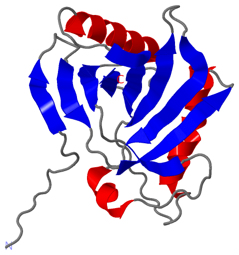
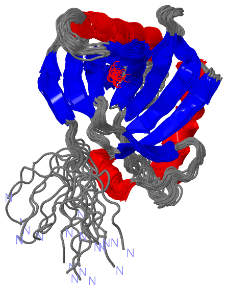
NMR StructureChain A from PDB Type:PROTEIN Length:184 aligned with HEBP1_MOUSE | Q9R257 from UniProtKB/Swiss-Prot Length:190 Alignment length:184 16 26 36 46 56 66 76 86 96 106 116 126 136 146 156 166 176 186 HEBP1_MOUSE 7 NSLFGSVETWPWQVLSTGGKEDVSYEERACEGGKFATVEVTDKPVDEALREAMPKIMKYVGGTNDKGVGMGMTVPVSFALFPNEDGSLQKKLKVWFRIPNQFQGSPPAPSDESVKIEEREGITVYSTQFGGYAKEADYVAHATQLRTTLEGTPATYQGDVYYCAGYDPPMKPYGRRNEVWLVKA 190 SCOP domains d2gova1 A:7-190 Heme-binding protein 1 SCOP domains CATH domains ---------------------------------------------------------------------------------------------------------------------------------------------------------------------------------------- CATH domains Pfam domains ---------------------------------------------------------------------------------------------------------------------------------------------------------------------------------------- Pfam domains SAPs(SNPs) ---------------------------------------------------------------------------------------------------------------------------------------------------------------------------------------- SAPs(SNPs) PROSITE ---------------------------------------------------------------------------------------------------------------------------------------------------------------------------------------- PROSITE Transcript ---------------------------------------------------------------------------------------------------------------------------------------------------------------------------------------- Transcript 2gov A 7 NSLFGSVETWPWQVLSTGGKEDVSYEERACEGGKFATVEVTDKPVDEALREAMPKIMKYVGGTNDKGVGMGMTVPVSFAVFPNEDGSLQKKLKVWFRIPNQFQGSPPAPSDESVKIEEREGITVYSTQFGGYAKEADYVAHATQLRTTLEGTPATYQGDVYYCAGYDPPMKPYGRRNEVWLVKA 190 16 26 36 46 56 66 76 86 96 106 116 126 136 146 156 166 176 186
|
||||||||||||||||||||
SCOP Domains (1, 1)
NMR Structure
 |
CATH Domains (0, 0)| (no "CATH Domain" information available for 2GOV) |
Pfam Domains (0, 0)| (no "Pfam Domain" information available for 2GOV) |
Gene Ontology (6, 6)|
NMR Structure(hide GO term definitions) Chain A (HEBP1_MOUSE | Q9R257)
|
||||||||||||||||||||||||||||||||||||||||||||||||||||||
Interactive Views
|
||||||||||||||||||||||||||||||||||||||||||||||||||||||||||||||||||||||||||||||||||||||||||||||||||||||||||||||||||||
Still Images
|
||||||||||||||||
Databases
|
||||||||||||||||||||||||||||||||||||||||||||||||||||||||||||||||||||||||||||||||||||||||||||||||||||||||||||||||||||||||||||||||||||||||||||||||||||||||||||||||
Analysis Tools
|
|||||||||||||||||||||||||||||||||||||||||||||||||||||||||||||
Entries Sharing at Least One Protein Chain (UniProt ID)
Related Entries Specified in the PDB File
|
|