| molecular function |
|---|
| | GO:0005524 | | ATP binding | | Interacting selectively and non-covalently with ATP, adenosine 5'-triphosphate, a universally important coenzyme and enzyme regulator. |
| | GO:0003824 | | catalytic activity | | Catalysis of a biochemical reaction at physiological temperatures. In biologically catalyzed reactions, the reactants are known as substrates, and the catalysts are naturally occurring macromolecular substances known as enzymes. Enzymes possess specific binding sites for substrates, and are usually composed wholly or largely of protein, but RNA that has catalytic activity (ribozyme) is often also regarded as enzymatic. |
| | GO:0004363 | | glutathione synthase activity | | Catalysis of the reaction: L-gamma-glutamyl-L-cysteine + ATP + glycine = ADP + glutathione + 2 H(+) + phosphate. |
| | GO:0016874 | | ligase activity | | Catalysis of the joining of two substances, or two groups within a single molecule, with the concomitant hydrolysis of the diphosphate bond in ATP or a similar triphosphate. |
| | GO:0000287 | | magnesium ion binding | | Interacting selectively and non-covalently with magnesium (Mg) ions. |
| | GO:0030145 | | manganese ion binding | | Interacting selectively and non-covalently with manganese (Mn) ions. |
| | GO:0046872 | | metal ion binding | | Interacting selectively and non-covalently with any metal ion. |
| | GO:0000166 | | nucleotide binding | | Interacting selectively and non-covalently with a nucleotide, any compound consisting of a nucleoside that is esterified with (ortho)phosphate or an oligophosphate at any hydroxyl group on the ribose or deoxyribose. |
| biological process |
|---|
| | GO:0006750 | | glutathione biosynthetic process | | The chemical reactions and pathways resulting in the formation of glutathione, the tripeptide glutamylcysteinylglycine, which acts as a coenzyme for some enzymes and as an antioxidant in the protection of sulfhydryl groups in enzymes and other proteins. |
| cellular component |
|---|
| | GO:0005829 | | cytosol | | The part of the cytoplasm that does not contain organelles but which does contain other particulate matter, such as protein complexes. |
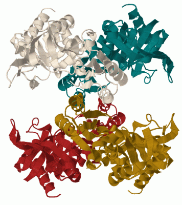
 Description
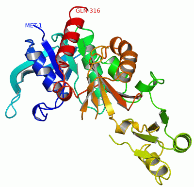