|
|
|
|
 Description
|
|
Compounds
|
||||||||||||||||||||||||||||||||||||||||
Chains, Units
Summary Information (see also Sequences/Alignments below) |
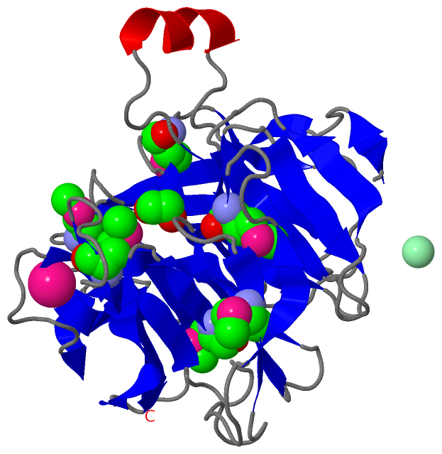
Ligands, Modified Residues, Ions (4, 9)| Asymmetric/Biological Unit (4, 9) |
Sites (3, 3)
Asymmetric Unit (3, 3)
|
SS Bonds (0, 0)| (no "SS Bond" information available for 2GHS) |
Cis Peptide Bonds (0, 0)| (no "Cis Peptide Bond" information available for 2GHS) |
SAPs(SNPs)/Variants (0, 0)| (no "SAP(SNP)/Variant" information available for 2GHS) |
PROSITE Motifs (0, 0)| (no "PROSITE Motif" information available for 2GHS) |
Exons (0, 0)| (no "Exon" information available for 2GHS) |
Sequences/Alignments
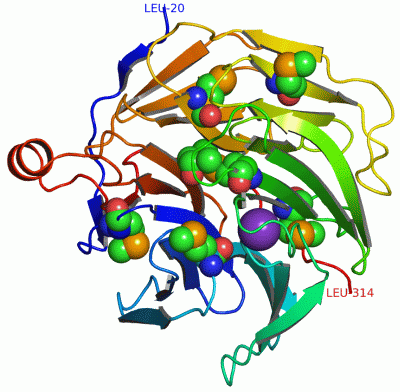
Asymmetric/Biological UnitChain A from PDB Type:PROTEIN Length:295 aligned with Q7D0W3_AGRFC | Q7D0W3 from UniProtKB/TrEMBL Length:295 Alignment length:295 10 20 30 40 50 60 70 80 90 100 110 120 130 140 150 160 170 180 190 200 210 220 230 240 250 260 270 280 290 Q7D0W3_AGRFC 1 MATVFPFAGRVLDETPMLLGEGPTFDPASGTAWWFNILERELHELHLASGRKTVHALPFMGSALAKISDSKQLIASDDGLFLRDTATGVLTLHAELESDLPGNRSNDGRMHPSGALWIGTMGRKAETGAGSIYHVAKGKVTKLFADISIPNSICFSPDGTTGYFVDTKVNRLMRVPLDARTGLPTGKAEVFIDSTGIKGGMDGSVCDAEGHIWNARWGEGAVDRYDTDGNHIARYEVPGKQTTCPAFIGPDASRLLVTSAREHLDDDAITANPQHGLTFELGIEVKGRFEPLYRL 295 SCOP domains d2ghsa1 A:20-314 Regucalcin SCOP domains CATH domains 2ghsA00 A:20-314 TolB, C-terminal domain CATH domains Pfam domains ------------------------------------------------------------------------------------------------------------------------------------------------------------------------------------------------------------------------------------------------------------------------------------------------------- Pfam domains SAPs(SNPs) ------------------------------------------------------------------------------------------------------------------------------------------------------------------------------------------------------------------------------------------------------------------------------------------------------- SAPs(SNPs) PROSITE ------------------------------------------------------------------------------------------------------------------------------------------------------------------------------------------------------------------------------------------------------------------------------------------------------- PROSITE Transcript ------------------------------------------------------------------------------------------------------------------------------------------------------------------------------------------------------------------------------------------------------------------------------------------------------- Transcript 2ghs A 20 LATVFPFAGRVLDETPmLLGEGPTFDPASGTAWWFNILERELHELHLASGRKTVHALPFmGSALAKISDSKQLIASDDGLFLRDTATGVLTLHAELESDLPGNRSNDGRmHPSGALWIGTmGRKAETGAGSIYHVAKGKVTKLFADISIPNSICFSPDGTTGYFVDTKVNRLmRVPLDARTGLPTGKAEVFIDSTGIKGGmDGSVCDAEGHIWNARWGEGAVDRYDTDGNHIARYEVPGKQTTCPAFIGPDASRLLVTSAREHLDDDAITANPQHGLTFELGIEVKGRFEPLYRL 314 29 | 39 49 59 69 79 89 99 109 119 129 139| 149 159 169 179 189 | 199 209 219| 229 239 249 259 269 279 289 299 309 36-MSE 79-MSE 129-MSE 140-MSE 192-MSE 220-MSE
|
||||||||||||||||||||
SCOP Domains (1, 1)
Asymmetric/Biological Unit

|
CATH Domains (1, 1)
Asymmetric/Biological Unit
|
Pfam Domains (0, 0)| (no "Pfam Domain" information available for 2GHS) |
Gene Ontology (0, 0)|
Asymmetric/Biological Unit(hide GO term definitions)
(no "Gene Ontology" information available for 2GHS)
|
Interactive Views
|
|||||||||||||||||||||||||||||||||||||||||||||||||||||||||||||||||||||||||||||||||||||||||||||||||||||||||||||||||||||||||||||||||||||||||||||||||||||||||
Still Images
|
||||||||||||||||
Databases
|
||||||||||||||||||||||||||||||||||||||||||||||||||||||||||||||||||||||||||||||||||||||||||||||||||||||||||||||||||||||||||||||||||||||||||||||||||||||||||||||||
Analysis Tools
|
|||||||||||||||||||||||||||||||||||||||||||||||||||||||||||||
Entries Sharing at Least One Protein Chain (UniProt ID)
Related Entries Specified in the PDB File
|
|