Show PDB file:   
         Plain Text   HTML   (compressed file size)
QuickSearch:   
by PDB,NDB,UniProt,PROSITE Code or Search Term(s)  
(-)Asym./Biol. Unit - manually
(-)Asym./Biol. Unit
(-)Asym./Biol. Unit - sites
collapse expand < >
Image Asym./Biol. Unit - manually
Asym./Biol. Unit - manually  (Jmol Viewer)
Image Asym./Biol. Unit
Asym./Biol. Unit  (Jmol Viewer)
Image Asym./Biol. Unit - sites
Asym./Biol. Unit - sites  (Jmol Viewer)

(-) Description

Title :  HUMAN DIPHOSPHOINOSITOL POLYPHOSPHATE PHOSPHOHYDROLASE 1
 
Authors :  B. M. Hallberg, P. Kursula, D. Ogg, C. Arrowsmith, H. Berglund, A. Edwards, M. Ehn, S. Flodin, S. Graslund, M. Hammarstrom, M. Hogbom, L. Holmberg-Schiavone, T. Kotenyova, P. Nilsson-Ehle, P. Nordlund, T. Nyman, J. Sagemark, P. Stenmark, M. Sundstrom, A. G. Thorsell, S. Van Den Berg, J. Weigelt, C. Persson, Structural Genomics Consortium (Sgc)
Date :  31 Jan 06  (Deposition) - 11 Mar 06  (Release) - 25 Aug 09  (Revision)
Method :  X-RAY DIFFRACTION
Resolution :  1.25
Chains :  Asym./Biol. Unit :  A
Keywords :  Diphosphoinositol Polyphosphate Phosphohydrolase, Nudix, Inositol Polyphosphate Metabolism, Structural Genomics, Structural Genomics Consortium, Sgc (Keyword Search: [Gene Ontology, PubMed, Web (Google))
 
Reference :  A. G. Thorsell, C. Persson, S. Graslund, M. Hammarstrom, R. D. Busam, B. M. Hallberg
Crystal Structure Of Human Diphosphoinositol Phosphatase 1
Proteins V. 77 242 2009
PubMed-ID: 19585659  |  Reference-DOI: 10.1002/PROT.22489
(for further references see the PDB file header)

(-) Compounds

Molecule 1 - DIPHOSPHOINOSITOL POLYPHOSPHATE PHOSPHOHYDROLASE 1
    ChainsA
    EC Number3.6.1.52, 3.6.1.-
    EngineeredYES
    Expression SystemESCHERICHIA COLI BL21(DE3)
    Expression System PlasmidPNIC-BSA4
    Expression System StrainBL21(DE3)
    Expression System Taxid469008
    Expression System Vector TypePLASMID
    FragmentRESIDUES 8-142
    GeneNUDT3, DIPP, DIPP1
    Organism CommonHUMAN
    Organism ScientificHOMO SAPIENS
    Organism Taxid9606
    SynonymDIPP-1, DIADENOSINE 5',5'''-P1,P6-HEXAPHOSPHATE HYDROLASE 1, NUCLEOSIDE DIPHOSPHATE-LINKED MOIETY X MOTIF 3, NUDIX MOTIF 3

 Structural Features

(-) Chains, Units

  1
Asymmetric/Biological Unit A

Summary Information (see also Sequences/Alignments below)

(-) Ligands, Modified Residues, Ions  (3, 3)

Asymmetric/Biological Unit (3, 3)
No.NameCountTypeFull Name
1CL1Ligand/IonCHLORIDE ION
2IHP1Ligand/IonINOSITOL HEXAKISPHOSPHATE
3SO41Ligand/IonSULFATE ION

(-) Sites  (3, 3)

Asymmetric Unit (3, 3)
No.NameEvidenceResiduesDescription
1AC1SOFTWARESER A:28 , SER A:30 , GLU A:31 , GLU A:32 , GLU A:33 , HOH A:4004BINDING SITE FOR RESIDUE CL A 2361
2AC2SOFTWAREARG A:20 , ASN A:86 , HIS A:91 , LYS A:133 , VAL A:135 , GLN A:136 , TYR A:139 , HOH A:4005 , HOH A:4010 , HOH A:4075 , HOH A:4237BINDING SITE FOR RESIDUE SO4 A 3001
3AC3SOFTWAREARG A:10 , LYS A:18 , ARG A:20 , SER A:39 , ARG A:41 , ARG A:89 , HIS A:91 , LYS A:133 , HOH A:4027 , HOH A:4031 , HOH A:4037 , HOH A:4049 , HOH A:4051 , HOH A:4053 , HOH A:4054 , HOH A:4060 , HOH A:4083 , HOH A:4089 , HOH A:4224BINDING SITE FOR RESIDUE IHP A 2360

(-) SS Bonds  (0, 0)

(no "SS Bond" information available for 2FVV)

(-) Cis Peptide Bonds  (0, 0)

(no "Cis Peptide Bond" information available for 2FVV)

 Sequence-Structure Mapping

(-) SAPs(SNPs)/Variants  (0, 0)

(no "SAP(SNP)/Variant" information available for 2FVV)

(-) PROSITE Motifs  (2, 2)

Asymmetric/Biological Unit (2, 2)
 PROSITEUniProtKBPDB
No.IDACDescriptionIDLocationCountLocation
1NUDIXPS51462 Nudix hydrolase domain profile.NUDT3_HUMAN17-142  1A:17-142
2NUDIX_BOXPS00893 Nudix box signature.NUDT3_HUMAN51-72  1A:51-72

(-) Exons   (0, 0)

(no "Exon" information available for 2FVV)

(-) Sequences/Alignments

Asymmetric/Biological Unit
   Reformat: Number of residues per line =  ('0' or empty: single-line sequence representation)
  Number of residues per labelling interval =   
  UniProt sequence: complete  aligned part    
   Show mapping: SCOP domains CATH domains Pfam domains Secondary structure (by author)
SAPs(SNPs) PROSITE motifs Exons
(details for a mapped element are shown in a popup box when the mouse pointer rests over it)
Chain A from PDB  Type:PROTEIN  Length:135
 aligned with NUDT3_HUMAN | O95989 from UniProtKB/Swiss-Prot  Length:172

    Alignment length:135
                                    17        27        37        47        57        67        77        87        97       107       117       127       137     
          NUDT3_HUMAN     8 QTRTYDGDGYKKRAACLCFRSESEEEVLLVSSSRHPDRWIVPGGGMEPEEEPSVAAVREVCEEAGVKGTLGRLVGIFENQERKHRTYVYVLIVTEVLEDWEDSVNIGRKREWFKIEDAIKVLQYHKPVQASYFET 142
               SCOP domains d2fvva1 A:8-142 Diphosphoinositol polyphosphate phosphohydrolase                                                                        SCOP domains
               CATH domains --------------------------------------------------------------------------------------------------------------------------------------- CATH domains
               Pfam domains --------------------------------------------------------------------------------------------------------------------------------------- Pfam domains
         Sec.struct. author ..........eeeeeeeee......eeeeee.......ee..eee.....hhhhhhhhhhhhhhheeeeeeeeeeeeee....eeeeeeeeeeeee...hhhhhhhh..eeeeehhhhhhhhh..hhhhhhhh.. Sec.struct. author
                 SAPs(SNPs) --------------------------------------------------------------------------------------------------------------------------------------- SAPs(SNPs)
                PROSITE (1) ---------NUDIX  PDB: A:17-142 UniProt: 17-142                                                                                           PROSITE (1)
                PROSITE (2) -------------------------------------------NUDIX_BOX  PDB: A:51-7---------------------------------------------------------------------- PROSITE (2)
                 Transcript --------------------------------------------------------------------------------------------------------------------------------------- Transcript
                 2fvv A   8 QTRTYDGDGYKKRAACLCFRSESEEEVLLVSSSRHPDRWIVPGGGMEPEEEPSVAAVREVCEEAGVKGTLGRLVGIFENQERKHRTYVYVLIVTEVLEDWEDSVNIGRKREWFKIEDAIKVLQYHKPVQASYFET 142
                                    17        27        37        47        57        67        77        87        97       107       117       127       137     

   Legend:   → Mismatch (orange background)
  - → Gap (green background, '-', border residues have a numbering label)
    → Modified Residue (blue background, lower-case, 'x' indicates undefined single-letter code, labelled with number + name)
  x → Chemical Group (purple background, 'x', labelled with number + name, e.g. ACE or NH2)
  extra numbering lines below/above indicate numbering irregularities and modified residue names etc., number ends below/above '|'

 Classification and Annotation

(-) SCOP Domains  (1, 1)

Asymmetric/Biological Unit

(-) CATH Domains  (0, 0)

(no "CATH Domain" information available for 2FVV)

(-) Pfam Domains  (0, 0)

(no "Pfam Domain" information available for 2FVV)

(-) Gene Ontology  (20, 20)

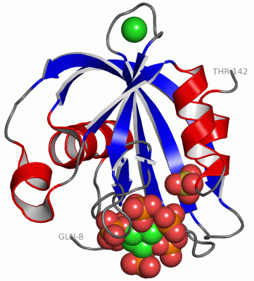
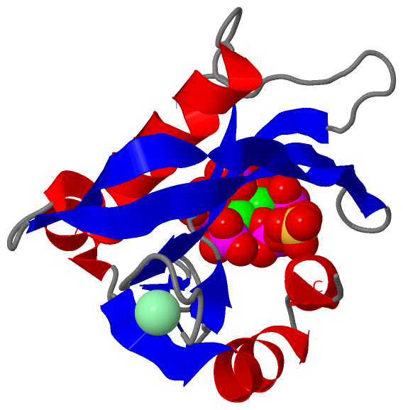
Asymmetric/Biological Unit(hide GO term definitions)
Chain A   (NUDT3_HUMAN | O95989)
molecular function
    GO:0008486    diphosphoinositol-polyphosphate diphosphatase activity    Catalysis of the reaction: diphospho-myo-inositol polyphosphate + H2O = myo-inositol polyphosphate + phosphate.
    GO:0016787    hydrolase activity    Catalysis of the hydrolysis of various bonds, e.g. C-O, C-N, C-C, phosphoric anhydride bonds, etc. Hydrolase is the systematic name for any enzyme of EC class 3.
    GO:0052841    inositol bisdiphosphate tetrakisphosphate diphosphatase activity    Catalysis of the reaction: inositol bisdiphosphate tetrakisphosphate + H2O = inositol diphosphate pentakisphosphate + phosphate.
    GO:0052842    inositol diphosphate pentakisphosphate diphosphatase activity    Catalysis of the reaction: inositol diphosphate pentakisphosphate + H2O = inositol hexakisphosphate + phosphate.
    GO:0052840    inositol diphosphate tetrakisphosphate diphosphatase activity    Catalysis of the reaction: inositol diphosphate tetrakisphosphate + H2O = inositol 1,3,4,5,6-pentakisphosphate + phosphate.
    GO:0052846    inositol-1,5-bisdiphosphate-2,3,4,6-tetrakisphosphate 1-diphosphatase activity    Catalysis of the reaction: 1,5-bisdiphosphoinositol-1D-myo-inositol 2,3,4,6-tetrakisphosphate + H2O = 1-diphospho-1D-myo-inositol 1,2,3,4,6-pentakisphosphate + phosphate + H+.
    GO:0052847    inositol-1,5-bisdiphosphate-2,3,4,6-tetrakisphosphate 5-diphosphatase activity    Catalysis of the reaction: 1,5-bisdiphosphoinositol-1D-myo-inositol 2,3,4,6-tetrakisphosphate + H2O = 1-diphospho-1D-myo-inositol 2,3,4,5,6-pentakisphosphate + phosphate + H+.
    GO:0052843    inositol-1-diphosphate-2,3,4,5,6-pentakisphosphate diphosphatase activity    Catalysis of the reaction: inositol 1-diphosphate 2,3,4,5,6-pentakisphosphate + H2O = inositol 1,2,3,4,5,6-hexakisphosphate + phosphate + 2 H+.
    GO:0052848    inositol-3,5-bisdiphosphate-2,3,4,6-tetrakisphosphate 5-diphosphatase activity    Catalysis of the reaction: 3,5-bisdiphosphoinositol-1D-myo-inositol 2,3,4,6-tetrakisphosphate + H2O = 3-diphospho-1D-myo-inositol 1,2,4,5,6-pentakisphosphate + phosphate + H+.
    GO:0052844    inositol-3-diphosphate-1,2,4,5,6-pentakisphosphate diphosphatase activity    Catalysis of the reaction: inositol 3-diphosphate 1,2,4,5,6-pentakisphosphate + H2O = inositol 1,2,3,4,5,6-hexakisphosphate + phosphate + 2 H+.
    GO:0052845    inositol-5-diphosphate-1,2,3,4,6-pentakisphosphate diphosphatase activity    Catalysis of the reaction: inositol 5-diphosphate 1,2,3,4,6-pentakisphosphate + H2O = inositol 1,2,3,4,5,6-hexakisphosphate + phosphate + 2 H+.
    GO:0000287    magnesium ion binding    Interacting selectively and non-covalently with magnesium (Mg) ions.
    GO:0046872    metal ion binding    Interacting selectively and non-covalently with any metal ion.
biological process
    GO:0007267    cell-cell signaling    Any process that mediates the transfer of information from one cell to another. This process includes signal transduction in the receiving cell and, where applicable, release of a ligand and any processes that actively facilitate its transport and presentation to the receiving cell. Examples include signaling via soluble ligands, via cell adhesion molecules and via gap junctions.
    GO:0015961    diadenosine polyphosphate catabolic process    The chemical reactions and pathways resulting in the breakdown of diadenosine polyphosphate, a derivative of the nucleoside adenosine with phosphate groups attached.
    GO:0071544    diphosphoinositol polyphosphate catabolic process    The chemical reactions and pathways resulting in the breakdown of a diphosphoinositol polyphosphate, 1,2,3,4,5,6-cyclohexanehexol with one or more diphosphate groups and multiple monophosphate groups attached.
    GO:0043647    inositol phosphate metabolic process    The chemical reactions and pathways involving inositol phosphate, 1,2,3,4,5,6-cyclohexanehexol, with one or more phosphate groups attached.
cellular component
    GO:0005737    cytoplasm    All of the contents of a cell excluding the plasma membrane and nucleus, but including other subcellular structures.
    GO:0005829    cytosol    The part of the cytoplasm that does not contain organelles but which does contain other particulate matter, such as protein complexes.
    GO:0070062    extracellular exosome    A vesicle that is released into the extracellular region by fusion of the limiting endosomal membrane of a multivesicular body with the plasma membrane. Extracellular exosomes, also simply called exosomes, have a diameter of about 40-100 nm.

 Visualization

(-) Interactive Views

Asymmetric/Biological Unit
  Complete Structure
    Jena3D(integrated viewing of ligand, site, SAP, PROSITE, SCOP information)
    WebMol | AstexViewer[tm]@PDBe
(Java Applets, require no local installation except for Java; loading may be slow)
    STRAP
(Java WebStart application, automatic local installation, requires Java; full application with system access!)
    RasMol
(require local installation)
    Molscript (VRML)
(requires installation of a VRML viewer; select preferred view via VRML and generate a mono or stereo PDF format file)
 
  Ligands, Modified Residues, Ions
    CL  [ RasMol | Jena3D ]  +environment [ RasMol | Jena3D ]
    IHP  [ RasMol | Jena3D ]  +environment [ RasMol | Jena3D ]
    SO4  [ RasMol | Jena3D ]  +environment [ RasMol | Jena3D ]
 
  Sites
    AC1  [ RasMol ]  +environment [ RasMol ]
    AC2  [ RasMol ]  +environment [ RasMol ]
    AC3  [ RasMol ]  +environment [ RasMol ]
 
  Cis Peptide Bonds
(no "Cis Peptide Bonds" information available for 2fvv)
 

(-) Still Images

Jmol
  protein: cartoon or spacefill or dots and stick; nucleic acid: cartoon and stick; ligands: spacefill; active site: stick
Molscript
  protein, nucleic acid: cartoon; ligands: spacefill; active site: ball and stick

 Databases and Analysis Tools

(-) Databases

Access by PDB/NDB ID
  2fvv
    Family and Domain InformationProDom | SYSTERS
    General Structural InformationGlycoscienceDB | MMDB | NDB | OCA | PDB | PDBe | PDBj | PDBsum | PDBWiki | PQS | PROTEOPEDIA
    Orientation in MembranesOPM
    Protein SurfaceSURFACE
    Secondary StructureDSSP (structure derived) | HSSP (homology derived)
    Structural GenomicsGeneCensus
    Structural NeighboursCE | VAST
    Structure ClassificationCATH | Dali | SCOP
    Validation and Original DataBMRB Data View | BMRB Restraints Grid | EDS | PROCHECK | RECOORD | WHAT_CHECK
 
Access by UniProt ID/Accession number
  NUDT3_HUMAN | O95989
    Comparative Protein Structure ModelsModBase
    Genomic InformationEnsembl
    Protein-protein InteractionDIP
    Sequence, Family and Domain InformationInterPro | Pfam | SMART | UniProtKB/SwissProt
 
Access by Enzyme Classificator   (EC Number)
  3.6.1.-
    General Enzyme InformationBRENDA | EC-PDB | Enzyme | IntEnz
    PathwayKEGG | MetaCyc
  3.6.1.52
    General Enzyme InformationBRENDA | EC-PDB | Enzyme | IntEnz
    PathwayKEGG | MetaCyc
 
Access by Disease Identifier   (MIM ID)
  (no 'MIM ID' available)
    Disease InformationOMIM
 
Access by GenAge ID
  (no 'GenAge ID' available)
    Age Related InformationGenAge

(-) Analysis Tools

Access by PDB/NDB ID
    Domain InformationXDom
    Interatomic Contacts of Structural UnitsCSU
    Ligand-protein ContactsLPC
    Protein CavitiescastP
    Sequence and Secondary StructurePDBCartoon
    Structure AlignmentSTRAP(Java WebStart application, automatic local installation, requires Java; full application with system access!)
    Structure and Sequence BrowserSTING
 
Access by UniProt ID/Accession number
  NUDT3_HUMAN | O95989
    Protein Disorder PredictionDisEMBL | FoldIndex | GLOBPLOT (for more information see DisProt)

 Related Entries

(-) Entries Sharing at Least One Protein Chain (UniProt ID)

UniProtKB/Swiss-Prot
        NUDT3_HUMAN | O959892q9p

(-) Related Entries Specified in the PDB File

(no "Related Entries Specified in the PDB File" available for 2FVV)