|
|
|
|
 Description
|
|
Compounds
|
||||||||||||||||||||||||||||||||||||||||||||
Chains, Units
Summary Information (see also Sequences/Alignments below) |
Ligands, Modified Residues, Ions (3, 5)| Asymmetric/Biological Unit (3, 5) |
Sites (2, 2)
Asymmetric Unit (2, 2)
|
SS Bonds (0, 0)| (no "SS Bond" information available for 2FSR) |
Cis Peptide Bonds (1, 1)
Asymmetric/Biological Unit
|
||||||||
SAPs(SNPs)/Variants (0, 0)| (no "SAP(SNP)/Variant" information available for 2FSR) |
PROSITE Motifs (0, 0)| (no "PROSITE Motif" information available for 2FSR) |
Exons (0, 0)| (no "Exon" information available for 2FSR) |
Sequences/Alignments
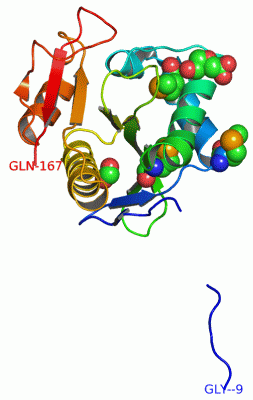
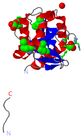
Asymmetric/Biological UnitChain A from PDB Type:PROTEIN Length:171 aligned with A9CHU9_AGRFC | A9CHU9 from UniProtKB/TrEMBL Length:171 Alignment length:171 1 | 6 16 26 36 46 56 66 76 86 96 106 116 126 136 146 156 166 A9CHU9_AGRFC - ----MNHSIPTLRTERLTLRPLAMADFPAYRDFMASPRSTGVGGPYDLPSTWGVFCHDLANWHFFGHGALMIDLGETGECIGQIGINHGPLFPEKELGWLLYEGHEGRGYAAEAAVALRDWAFETLNLPTLVSYVSPQNRKSAAVAERIGGTLDPLAPRSDPEDLVYRYHQ 167 SCOP domains -------d2fsra1 A:4-167 Probable acetyltranferase Atu2435 SCOP domains CATH domains 2fsrA00 A:-9-167 [code=3.40.630.30, no name defined] CATH domains Pfam domains --------------------------------------------------------------------------------------------------------------------------------------------------------------------------- Pfam domains SAPs(SNPs) --------------------------------------------------------------------------------------------------------------------------------------------------------------------------- SAPs(SNPs) PROSITE --------------------------------------------------------------------------------------------------------------------------------------------------------------------------- PROSITE Transcript --------------------------------------------------------------------------------------------------------------------------------------------------------------------------- Transcript 2fsr A -9 GRENLYFSIPTLRTERLTLRPLAmADFPAYRDFmASPRSTGVGGPYDLPSTWGVFCHDLANWHFFGHGALmIDLGETGECIGQIGINHGPLFPEKELGWLLYEGHEGRGYAAEAAVALRDWAFETLNLPTLVSYVSPQNRKSAAVAERIGGTLDPLAPRSDPEDLVYRYHQ 167 || 6 16 | 26 | 36 46 56 66| 76 86 96 106 116 126 136 146 156 166 -3| 20-MSE 30-MSE 67-MSE 4
|
||||||||||||||||||||
SCOP Domains (1, 1)
Asymmetric/Biological Unit
 |
CATH Domains (1, 1)
Asymmetric/Biological Unit
|
Pfam Domains (0, 0)| (no "Pfam Domain" information available for 2FSR) |
Gene Ontology (1, 1)|
Asymmetric/Biological Unit(hide GO term definitions) Chain A (A9CHU9_AGRFC | A9CHU9)
|
||||||||||||
Interactive Views
|
||||||||||||||||||||||||||||||||||||||||||||||||||||||||||||||||||||||||||||||||||||||||||||||||||||||||||||||||||||||||||||||||||||||||||||
Still Images
|
||||||||||||||||
Databases
|
||||||||||||||||||||||||||||||||||||||||||||||||||||||||||||||||||||||||||||||||||||||||||||||||||||||||||||||||||||||||||||||||||||||||||||||||||||||||||||||||
Analysis Tools
|
|||||||||||||||||||||||||||||||||||||||||||||||||||||||||||||
Entries Sharing at Least One Protein Chain (UniProt ID)
Related Entries Specified in the PDB File
|
|