| molecular function |
|---|
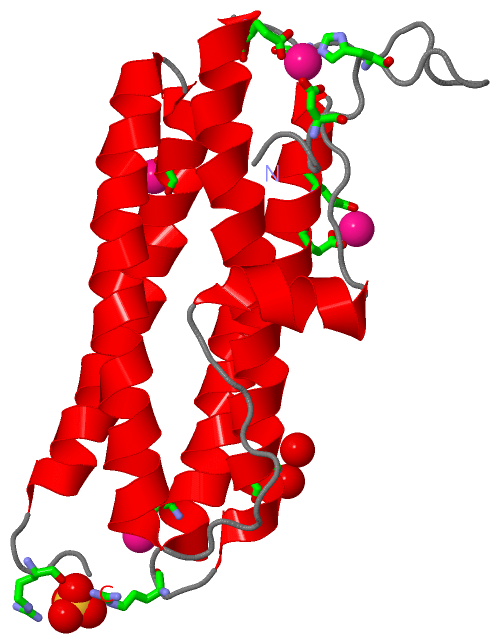
| | GO:0003677 | | DNA binding | | Any molecular function by which a gene product interacts selectively and non-covalently with DNA (deoxyribonucleic acid). |
| | GO:0008199 | | ferric iron binding | | Interacting selectively and non-covalently with ferric iron, Fe(III). |
| | GO:0046872 | | metal ion binding | | Interacting selectively and non-covalently with any metal ion. |
| | GO:0016491 | | oxidoreductase activity | | Catalysis of an oxidation-reduction (redox) reaction, a reversible chemical reaction in which the oxidation state of an atom or atoms within a molecule is altered. One substrate acts as a hydrogen or electron donor and becomes oxidized, while the other acts as hydrogen or electron acceptor and becomes reduced. |
| | GO:0016722 | | oxidoreductase activity, oxidizing metal ions | | Catalysis of an oxidation-reduction in which the oxidation state of metal ion is altered. |
| biological process |
|---|
| | GO:0006879 | | cellular iron ion homeostasis | | Any process involved in the maintenance of an internal steady state of iron ions at the level of a cell. |
| | GO:0055114 | | oxidation-reduction process | | A metabolic process that results in the removal or addition of one or more electrons to or from a substance, with or without the concomitant removal or addition of a proton or protons. |
| | GO:0006950 | | response to stress | | Any process that results in a change in state or activity of a cell or an organism (in terms of movement, secretion, enzyme production, gene expression, etc.) as a result of a disturbance in organismal or cellular homeostasis, usually, but not necessarily, exogenous (e.g. temperature, humidity, ionizing radiation). |
| cellular component |
|---|
| | GO:0005737 | | cytoplasm | | All of the contents of a cell excluding the plasma membrane and nucleus, but including other subcellular structures. |
| | GO:0009295 | | nucleoid | | The region of a virus, bacterial cell, mitochondrion or chloroplast to which the nucleic acid is confined. |
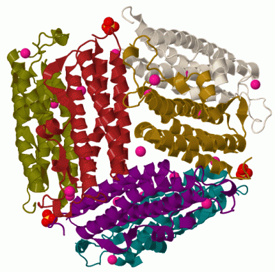
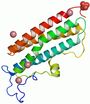
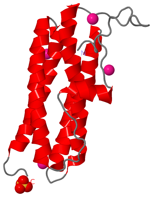
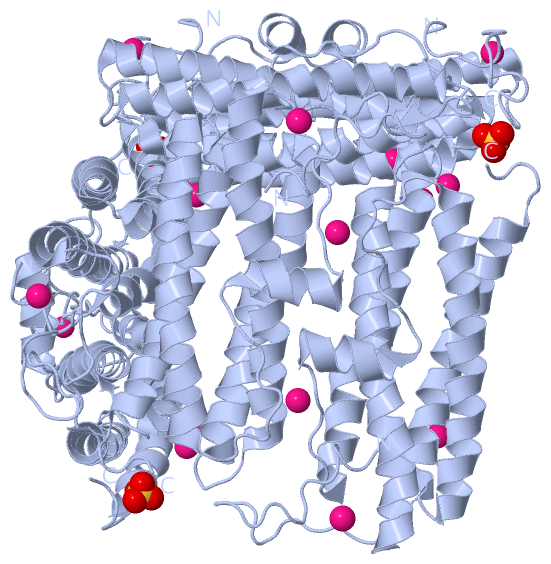
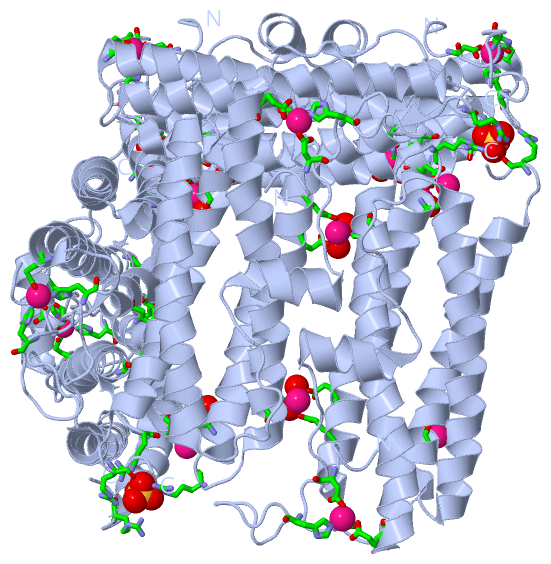
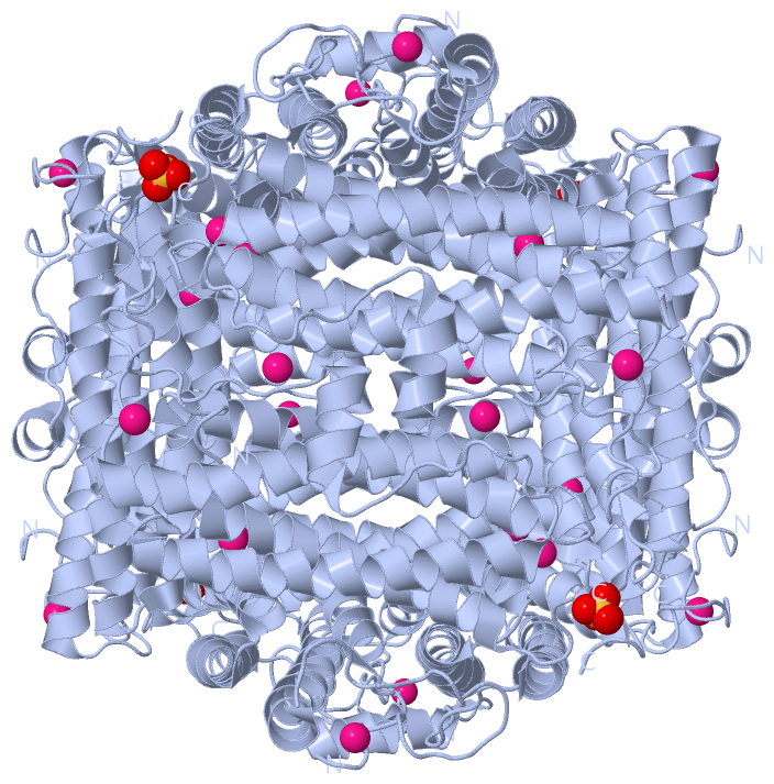
 Description