Show PDB file:   
         Plain Text   HTML   (compressed file size)
QuickSearch:   
by PDB,NDB,UniProt,PROSITE Code or Search Term(s)  
(-)Asym./Biol. Unit
(-)Asym./Biol. Unit - sites
collapse expand < >
Image Asym./Biol. Unit
Asym./Biol. Unit  (Jmol Viewer)
Image Asym./Biol. Unit - sites
Asym./Biol. Unit - sites  (Jmol Viewer)

(-) Description

Title :  CRYSTAL STRUCTURE OF A PUTATIVE DNA DAMAGE-INDUCABLE (DINB) PROTEIN (BH3987) FROM BACILLUS HALODURANS AT 1.42 A RESOLUTION
 
Authors :  Joint Center For Structural Genomics (Jcsg)
Date :  15 Nov 05  (Deposition) - 20 Dec 05  (Release) - 13 Jul 11  (Revision)
Method :  X-RAY DIFFRACTION
Resolution :  1.42
Chains :  Asym./Biol. Unit :  A,B
Keywords :  Putative Dna Damage-Inducable (Dinb) Protein, Structural Genomics, Joint Center For Structural Genomics, Jcsg, Protein Structure Initiative, Psi-2, Metal Binding Protein (Keyword Search: [Gene Ontology, PubMed, Web (Google))
 
Reference :  Joint Center For Structural Genomics (Jcsg)
Crystal Structure Of Bh3987 From Bacillus Halodurans At 1. 4 A Resolution
To Be Published
PubMed: search

(-) Compounds

Molecule 1 - BH3987
    ChainsA, B
    EngineeredYES
    Expression SystemESCHERICHIA COLI
    Expression System Taxid562
    Expression System Vector TypePLASMID
    Gene10176612
    Organism ScientificBACILLUS HALODURANS
    Organism Taxid86665

 Structural Features

(-) Chains, Units

  12
Asymmetric/Biological Unit AB

Summary Information (see also Sequences/Alignments below)

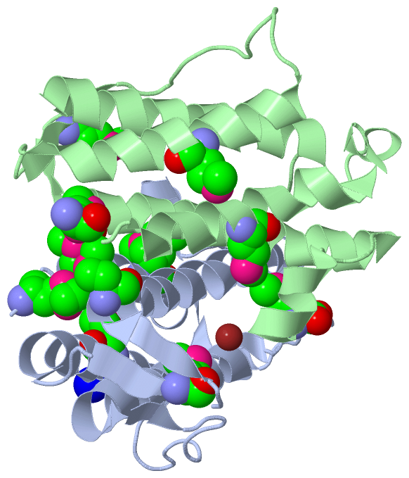
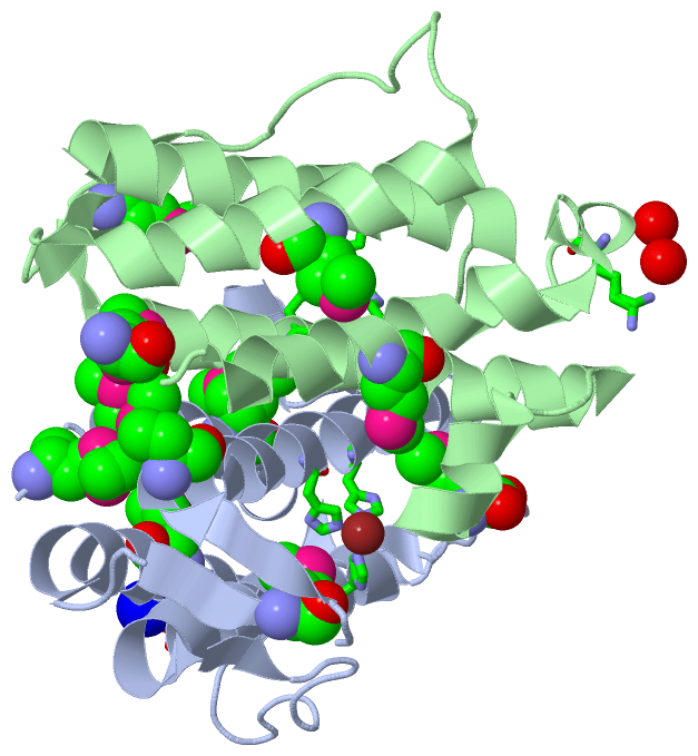
(-) Ligands, Modified Residues, Ions  (3, 14)

Asymmetric/Biological Unit (3, 14)
No.NameCountTypeFull Name
1MSE11Mod. Amino AcidSELENOMETHIONINE
2NA1Ligand/IonSODIUM ION
3NI2Ligand/IonNICKEL (II) ION

(-) Sites  (3, 3)

Asymmetric Unit (3, 3)
No.NameEvidenceResiduesDescription
1AC1SOFTWAREHIS B:42 , HIS B:114 , HIS B:118 , HOH B:192 , HOH B:195 , HOH B:196BINDING SITE FOR RESIDUE NI B 144
2AC2SOFTWAREHIS A:42 , HIS A:114 , HIS A:118 , HOH A:181 , HOH A:195 , HOH A:220BINDING SITE FOR RESIDUE NI A 144
3AC3SOFTWAREASP A:74 , HOH A:368 , LYS B:24 , HOH B:232 , HOH B:235BINDING SITE FOR RESIDUE NA A 145

(-) SS Bonds  (0, 0)

(no "SS Bond" information available for 2F22)

(-) Cis Peptide Bonds  (2, 2)

Asymmetric/Biological Unit
No.Residues
1Phe A:61 -Pro A:62
2Phe B:61 -Pro B:62

 Sequence-Structure Mapping

(-) SAPs(SNPs)/Variants  (0, 0)

(no "SAP(SNP)/Variant" information available for 2F22)

(-) PROSITE Motifs  (0, 0)

(no "PROSITE Motif" information available for 2F22)

(-) Exons   (0, 0)

(no "Exon" information available for 2F22)

(-) Sequences/Alignments

Asymmetric/Biological Unit
   Reformat: Number of residues per line =  ('0' or empty: single-line sequence representation)
  Number of residues per labelling interval =   
  UniProt sequence: complete  aligned part    
   Show mapping: SCOP domains CATH domains Pfam domains Secondary structure (by author)
SAPs(SNPs) PROSITE motifs Exons
(details for a mapped element are shown in a popup box when the mouse pointer rests over it)
Chain A from PDB  Type:PROTEIN  Length:142
 aligned with Q9RC77_BACHO | Q9RC77 from UniProtKB/TrEMBL  Length:143

    Alignment length:142
                             1                                                                                                                                            
                             |       9        19        29        39        49        59        69        79        89        99       109       119       129       139  
         Q9RC77_BACHO     - -MDTNGVLYAANMTNALAKEIPESKWDIQLIPELGTLRKLFIHIVRVRDVYRDGLKTGSIKFPGRLASDEHRLLDELERSMEELVFEFKQTTFNSIKMGENYLSIMELLGTVIQHEGIHQGQYYVALKQSGINLPKQWVQDW 141
               SCOP domains -d2f22a1 A:1-141 Hypothetical protein BH3987                                                                                                   SCOP domains
               CATH domains ---------------------------------------------------------------------------------------------------------------------------------------------- CATH domains
               Pfam domains ---------------------------------------------------------------------------------------------------------------------------------------------- Pfam domains
         Sec.struct. author ...hhhhhhhhhhhhhhhhhhhhhhh.........hhhhhhhhhhhhhhhhhhhhhhhee...........hhhhhhhhhhhhhhhhhhhh...eeee..eeeehhhhhhhhhhhhhhhhhhhhhhhhhhh...hhhhhhhh Sec.struct. author
                 SAPs(SNPs) ---------------------------------------------------------------------------------------------------------------------------------------------- SAPs(SNPs)
                    PROSITE ---------------------------------------------------------------------------------------------------------------------------------------------- PROSITE
                 Transcript ---------------------------------------------------------------------------------------------------------------------------------------------- Transcript
                 2f22 A   0 GmDTNGVLYAANmTNALAKEIPESKWDIQLIPELGTLRKLFIHIVRVRDVYRDGLKTGSIKFPGRLASDEHRLLDELERSmEELVFEFKQTTFNSIKmGENYLSImELLGTVIQHEGIHQGQYYVALKQSGINLPKQWVQDW 141
                             |       9  |     19        29        39        49        59        69        79|       89       |99     | 109       119       129       139  
                             |         12-MSE                                                              80-MSE           97-MSE 105-MSE                                
                             1-MSE                                                                                                                                        

Chain B from PDB  Type:PROTEIN  Length:143
 aligned with Q9RC77_BACHO | Q9RC77 from UniProtKB/TrEMBL  Length:143

    Alignment length:143
                                    10        20        30        40        50        60        70        80        90       100       110       120       130       140   
         Q9RC77_BACHO     1 MDTNGVLYAANMTNALAKEIPESKWDIQLIPELGTLRKLFIHIVRVRDVYRDGLKTGSIKFPGRLASDEHRLLDELERSMEELVFEFKQTTFNSIKMGENYLSIMELLGTVIQHEGIHQGQYYVALKQSGINLPKQWVQDWHM 143
               SCOP domains d2f22b_ B: Hypothetical protein BH3987                                                                                                          SCOP domains
               CATH domains ----------------------------------------------------------------------------------------------------------------------------------------------- CATH domains
               Pfam domains ----------------------------------------------------------------------------------------------------------------------------------------------- Pfam domains
         Sec.struct. author ..hhhhhhhhhhhhhhhhhhhhhhh.........hhhhhhhhhhhhhhhhhhhhhhhee...........hhhhhhhhhhhhhhhhhhhh...eeee..eeeehhhhhhhhhhhhhhhhhhhhhhhhhhh...hhhhhhhhh. Sec.struct. author
                 SAPs(SNPs) ----------------------------------------------------------------------------------------------------------------------------------------------- SAPs(SNPs)
                    PROSITE ----------------------------------------------------------------------------------------------------------------------------------------------- PROSITE
                 Transcript ----------------------------------------------------------------------------------------------------------------------------------------------- Transcript
                 2f22 B   1 mDTNGVLYAANmTNALAKEIPESKWDIQLIPELGTLRKLFIHIVRVRDVYRDGLKTGSIKFPGRLASDEHRLLDELERSmEELVFEFKQTTFNSIKmGENYLSImELLGTVIQHEGIHQGQYYVALKQSGINLPKQWVQDWHm 143
                            |       10 |      20        30        40        50        60        70        80        90      |100    |  110       120       130       140  |
                            1-MSE     12-MSE                                                              80-MSE           97-MSE 105-MSE                               143-MSE

   Legend:   → Mismatch (orange background)
  - → Gap (green background, '-', border residues have a numbering label)
    → Modified Residue (blue background, lower-case, 'x' indicates undefined single-letter code, labelled with number + name)
  x → Chemical Group (purple background, 'x', labelled with number + name, e.g. ACE or NH2)
  extra numbering lines below/above indicate numbering irregularities and modified residue names etc., number ends below/above '|'

 Classification and Annotation

(-) SCOP Domains  (1, 2)

Asymmetric/Biological Unit

(-) CATH Domains  (0, 0)

(no "CATH Domain" information available for 2F22)

(-) Pfam Domains  (0, 0)

(no "Pfam Domain" information available for 2F22)

(-) Gene Ontology  (0, 0)

Asymmetric/Biological Unit(hide GO term definitions)
    (no "Gene Ontology" information available for 2F22)

 Visualization

(-) Interactive Views

Asymmetric/Biological Unit
  Complete Structure
    Jena3D(integrated viewing of ligand, site, SAP, PROSITE, SCOP information)
    WebMol | AstexViewer[tm]@PDBe
(Java Applets, require no local installation except for Java; loading may be slow)
    STRAP
(Java WebStart application, automatic local installation, requires Java; full application with system access!)
    RasMol
(require local installation)
    Molscript (VRML)
(requires installation of a VRML viewer; select preferred view via VRML and generate a mono or stereo PDF format file)
 
  Ligands, Modified Residues, Ions
    MSE  [ RasMol | Jena3D ]  +environment [ RasMol | Jena3D ]
    NA  [ RasMol | Jena3D ]  +environment [ RasMol | Jena3D ]
    NI  [ RasMol | Jena3D ]  +environment [ RasMol | Jena3D ]
 
  Sites
    AC1  [ RasMol ]  +environment [ RasMol ]
    AC2  [ RasMol ]  +environment [ RasMol ]
    AC3  [ RasMol ]  +environment [ RasMol ]
 
  Cis Peptide Bonds
    Phe A:61 - Pro A:62   [ RasMol ]  
    Phe B:61 - Pro B:62   [ RasMol ]  
 

(-) Still Images

Jmol
  protein: cartoon or spacefill or dots and stick; nucleic acid: cartoon and stick; ligands: spacefill; active site: stick
Molscript
  protein, nucleic acid: cartoon; ligands: spacefill; active site: ball and stick

 Databases and Analysis Tools

(-) Databases

Access by PDB/NDB ID
  2f22
    Family and Domain InformationProDom | SYSTERS
    General Structural InformationGlycoscienceDB | MMDB | NDB | OCA | PDB | PDBe | PDBj | PDBsum | PDBWiki | PQS | PROTEOPEDIA
    Orientation in MembranesOPM
    Protein SurfaceSURFACE
    Secondary StructureDSSP (structure derived) | HSSP (homology derived)
    Structural GenomicsGeneCensus
    Structural NeighboursCE | VAST
    Structure ClassificationCATH | Dali | SCOP
    Validation and Original DataBMRB Data View | BMRB Restraints Grid | EDS | PROCHECK | RECOORD | WHAT_CHECK
 
Access by UniProt ID/Accession number
  Q9RC77_BACHO | Q9RC77
    Comparative Protein Structure ModelsModBase
    Genomic InformationEnsembl
    Protein-protein InteractionDIP
    Sequence, Family and Domain InformationInterPro | Pfam | SMART | UniProtKB/TrEMBL
 
Access by Enzyme Classificator   (EC Number)
  (no 'Enzyme Classificator' available)
    General Enzyme InformationBRENDA | EC-PDB | Enzyme | IntEnz
    PathwayKEGG | MetaCyc
 
Access by Disease Identifier   (MIM ID)
  (no 'MIM ID' available)
    Disease InformationOMIM
 
Access by GenAge ID
  (no 'GenAge ID' available)
    Age Related InformationGenAge

(-) Analysis Tools

Access by PDB/NDB ID
    Domain InformationXDom
    Interatomic Contacts of Structural UnitsCSU
    Ligand-protein ContactsLPC
    Protein CavitiescastP
    Sequence and Secondary StructurePDBCartoon
    Structure AlignmentSTRAP(Java WebStart application, automatic local installation, requires Java; full application with system access!)
    Structure and Sequence BrowserSTING
 
Access by UniProt ID/Accession number
  Q9RC77_BACHO | Q9RC77
    Protein Disorder PredictionDisEMBL | FoldIndex | GLOBPLOT (for more information see DisProt)

 Related Entries

(-) Entries Sharing at Least One Protein Chain (UniProt ID)

(no "Entries Sharing at Least One Protein Chain" available for 2F22)

(-) Related Entries Specified in the PDB File

(no "Related Entries Specified in the PDB File" available for 2F22)