Show PDB file:   
         Plain Text   HTML   (compressed file size)
QuickSearch:   
by PDB,NDB,UniProt,PROSITE Code or Search Term(s)  
(-)NMR Structure - manually
(-)NMR Structure - model 1
(-)NMR Structure - all models
collapse expand < >
Image NMR Structure - manually
NMR Structure - manually  (Jmol Viewer)
Image NMR Structure - model 1
NMR Structure - model 1  (Jmol Viewer)
Image NMR Structure - all models
NMR Structure - all models  (Jmol Viewer)

(-) Description

Title :  USE OF RESTRAINED MOLECULAR DYNAMICS IN WATER TO DETERMINE THREE-DIMENSIONAL PROTEIN STRUCTURE: PREDICTION OF THE THREE-DIMENSIONAL STRUCTURE OF ECBALLIUM ELATERIUM TRYPSIN INHIBITOR II
 
Authors :  A. Heitz, L. Chiche, D. Le-Nguyen, B. Castro
Date :  15 Jul 91  (Deposition) - 15 Oct 91  (Release) - 24 Feb 09  (Revision)
Method :  SOLUTION NMR
Resolution :  NOT APPLICABLE
Chains :  NMR Structure  :  A
Keywords :  Protein Inhibitor (Keyword Search: [Gene Ontology, PubMed, Web (Google))
 
Reference :  L. Chiche, C. Gaboriaud, A. Heitz, J. P. Mornon, B. Castro, P. A. Kollman
Use Of Restrained Molecular Dynamics In Water To Determine Three-Dimensional Protein Structure: Prediction Of The Three-Dimensional Structure Of Ecballium Elaterium Trypsin Inhibitor Ii.
Proteins V. 6 405 1989
PubMed-ID: 2622910  |  Reference-DOI: 10.1002/PROT.340060407
(for further references see the PDB file header)

(-) Compounds

Molecule 1 - TRYPSIN INHIBITOR II
    ChainsA
    EngineeredYES
    Organism CommonJUMPING CUCUMBER
    Organism ScientificECBALLIUM ELATERIUM
    Organism Taxid3679

 Structural Features

(-) Chains, Units

  
NMR Structure 

Summary Information (see also Sequences/Alignments below)

(-) Ligands, Modified Residues, Ions  (0, 0)

(no "Ligand,Modified Residues,Ions" information available for 2ETI)

(-) Sites  (0, 0)

(no "Site" information available for 2ETI)
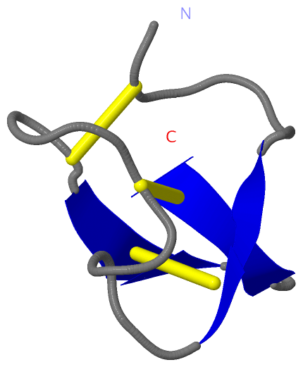
(-) SS Bonds  (3, 3)

NMR Structure
No.Residues
1A:2 -A:19
2A:9 -A:21
3A:15 -A:27

(-) Cis Peptide Bonds  (0, 0)

(no "Cis Peptide Bond" information available for 2ETI)

 Sequence-Structure Mapping

(-) SAPs(SNPs)/Variants  (0, 0)

(no "SAP(SNP)/Variant" information available for 2ETI)

(-) PROSITE Motifs  (1, 1)

NMR Structure (1, 1)
 PROSITEUniProtKBPDB
No.IDACDescriptionIDLocationCountLocation
1SQUASH_INHIBITORPS00286 Squash family of serine protease inhibitors signature.ITR2_ECBEL2-21  1A:2-21

(-) Exons   (0, 0)

(no "Exon" information available for 2ETI)

(-) Sequences/Alignments

NMR Structure
   Reformat: Number of residues per line =  ('0' or empty: single-line sequence representation)
  Number of residues per labelling interval =   
  UniProt sequence: complete  aligned part    
   Show mapping: SCOP domains CATH domains Pfam domains Secondary structure (by author)
SAPs(SNPs) PROSITE motifs Exons
(details for a mapped element are shown in a popup box when the mouse pointer rests over it)
Chain A from PDB  Type:PROTEIN  Length:28
 aligned with ITR2_ECBEL | P12071 from UniProtKB/Swiss-Prot  Length:30

    Alignment length:28
                                    10        20        
            ITR2_ECBEL    1 GCPRILMRCKQDSDCLAGCVCGPNGFCG 28
               SCOP domains d2etia_ A: Trypsin inhibitor SCOP domains
               CATH domains ---------------------------- CATH domains
               Pfam domains ---------------------------- Pfam domains
         Sec.struct. author ......eee..........eee..eeee Sec.struct. author
                 SAPs(SNPs) ---------------------------- SAPs(SNPs)
                    PROSITE -SQUASH_INHIBITOR    ------- PROSITE
                 Transcript ---------------------------- Transcript
                  2eti A  1 GCPRILMRCKQDSDCLAGCVCGPNGFCG 28
                                    10        20        

   Legend:   → Mismatch (orange background)
  - → Gap (green background, '-', border residues have a numbering label)
    → Modified Residue (blue background, lower-case, 'x' indicates undefined single-letter code, labelled with number + name)
  x → Chemical Group (purple background, 'x', labelled with number + name, e.g. ACE or NH2)
  extra numbering lines below/above indicate numbering irregularities and modified residue names etc., number ends below/above '|'

 Classification and Annotation

(-) SCOP Domains  (1, 1)

NMR Structure

(-) CATH Domains  (0, 0)

(no "CATH Domain" information available for 2ETI)

(-) Pfam Domains  (0, 0)

(no "Pfam Domain" information available for 2ETI)

(-) Gene Ontology  (6, 6)

NMR Structure(hide GO term definitions)
Chain A   (ITR2_ECBEL | P12071)
molecular function
    GO:0004866    endopeptidase inhibitor activity    Stops, prevents or reduces the activity of an endopeptidase, any enzyme that hydrolyzes nonterminal peptide bonds in polypeptides.
    GO:0030414    peptidase inhibitor activity    Stops, prevents or reduces the activity of a peptidase, any enzyme that catalyzes the hydrolysis peptide bonds.
    GO:0004867    serine-type endopeptidase inhibitor activity    Stops, prevents or reduces the activity of serine-type endopeptidases, enzymes that catalyze the hydrolysis of nonterminal peptide bonds in a polypeptide chain; a serine residue (and a histidine residue) are at the active center of the enzyme.
biological process
    GO:0010951    negative regulation of endopeptidase activity    Any process that decreases the frequency, rate or extent of endopeptidase activity, the endohydrolysis of peptide bonds within proteins.
    GO:0010466    negative regulation of peptidase activity    Any process that stops or reduces the rate of peptidase activity, the hydrolysis of peptide bonds within proteins.
cellular component
    GO:0005576    extracellular region    The space external to the outermost structure of a cell. For cells without external protective or external encapsulating structures this refers to space outside of the plasma membrane. This term covers the host cell environment outside an intracellular parasite.

 Visualization

(-) Interactive Views

NMR Structure
  Complete Structure
    Jena3D(integrated viewing of ligand, site, SAP, PROSITE, SCOP information)
    WebMol | AstexViewer[tm]@PDBe
(Java Applets, require no local installation except for Java; loading may be slow)
    STRAP
(Java WebStart application, automatic local installation, requires Java; full application with system access!)
    RasMol
(require local installation)
    Molscript (VRML)
(requires installation of a VRML viewer; select preferred view via VRML and generate a mono or stereo PDF format file)
 
  Ligands, Modified Residues, Ions
(no "Ligands, Modified Residues, Ions" information available for 2eti)
 
  Sites
(no "Sites" information available for 2eti)
 
  Cis Peptide Bonds
(no "Cis Peptide Bonds" information available for 2eti)
 

(-) Still Images

Jmol
  protein: cartoon or spacefill or dots and stick; nucleic acid: cartoon and stick; ligands: spacefill; active site: stick
Molscript
  protein, nucleic acid: cartoon; ligands: spacefill; active site: ball and stick
Midas
  ribbon, secondary structure
  sticks
  spacefill
Prepi
  C alpha wire, SS bonds, sequence
  sticks, SS bonds
  ribbon, secondary structure, SS bonds, sticks
  ribbon,secondary structure, SS bonds
Setor
  ribbon, SS bonds, numbering
  ribbon, secondary structure, SS bonds, numbering
  sticks, numbering
Distance Plot
  representative atom: CA

 Databases and Analysis Tools

(-) Databases

Access by PDB/NDB ID
  2eti
    Family and Domain InformationProDom | SYSTERS
    General Structural InformationGlycoscienceDB | MMDB | NDB | OCA | PDB | PDBe | PDBj | PDBsum | PDBWiki | PQS | PROTEOPEDIA
    Orientation in MembranesOPM
    Protein SurfaceSURFACE
    Secondary StructureDSSP (structure derived) | HSSP (homology derived)
    Structural GenomicsGeneCensus
    Structural NeighboursCE | VAST
    Structure ClassificationCATH | Dali | SCOP
    Validation and Original DataBMRB Data View | BMRB Restraints Grid | EDS | PROCHECK | RECOORD | WHAT_CHECK
 
Access by UniProt ID/Accession number
  ITR2_ECBEL | P12071
    Comparative Protein Structure ModelsModBase
    Genomic InformationEnsembl
    Protein-protein InteractionDIP
    Sequence, Family and Domain InformationInterPro | Pfam | SMART | UniProtKB/SwissProt
 
Access by Enzyme Classificator   (EC Number)
  (no 'Enzyme Classificator' available)
    General Enzyme InformationBRENDA | EC-PDB | Enzyme | IntEnz
    PathwayKEGG | MetaCyc
 
Access by Disease Identifier   (MIM ID)
  (no 'MIM ID' available)
    Disease InformationOMIM
 
Access by GenAge ID
  (no 'GenAge ID' available)
    Age Related InformationGenAge

(-) Analysis Tools

Access by PDB/NDB ID
    Domain InformationXDom
    Interatomic Contacts of Structural UnitsCSU
    Ligand-protein ContactsLPC
    Protein CavitiescastP
    Sequence and Secondary StructurePDBCartoon
    Structure AlignmentSTRAP(Java WebStart application, automatic local installation, requires Java; full application with system access!)
    Structure and Sequence BrowserSTING
 
Access by UniProt ID/Accession number
  ITR2_ECBEL | P12071
    Protein Disorder PredictionDisEMBL | FoldIndex | GLOBPLOT (for more information see DisProt)

 Related Entries

(-) Entries Sharing at Least One Protein Chain (UniProt ID)

UniProtKB/Swiss-Prot
        ITR2_ECBEL | P120711h9h 1h9i 1w7z 2c4b 2it7 2let

(-) Related Entries Specified in the PDB File

(no "Related Entries Specified in the PDB File" available for 2ETI)