|
|
|
|
 Description
|
|
Compounds
|
||||||||||||||||||||||||||||||||||||||||||||||||
Chains, Units
Summary Information (see also Sequences/Alignments below) |
Ligands, Modified Residues, Ions (1, 1)
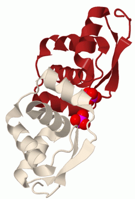
Asymmetric Unit (1, 1)
|
Sites (1, 1)
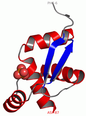
Asymmetric Unit (1, 1)
|
SS Bonds (0, 0)| (no "SS Bond" information available for 2EFV) |
Cis Peptide Bonds (0, 0)| (no "Cis Peptide Bond" information available for 2EFV) |
SAPs(SNPs)/Variants (0, 0)| (no "SAP(SNP)/Variant" information available for 2EFV) |
PROSITE Motifs (0, 0)| (no "PROSITE Motif" information available for 2EFV) |
Exons (0, 0)| (no "Exon" information available for 2EFV) |
Sequences/Alignments
Asymmetric UnitChain A from PDB Type:PROTEIN Length:82 aligned with Y366_METJA | Q57812 from UniProtKB/Swiss-Prot Length:92 Alignment length:82 15 25 35 45 55 65 75 85 Y366_METJA 6 FMKEKKRATFYLYKNIDGRKLRYLLHKLENVENVDIDTLRRAIEAEKKYKRSITLTEEEEVIIQRLGKSANLLLNCELVKLD 87 SCOP domains d2efva1 A:6-87 Uncharacterized protein MJ0366 SCOP domains CATH domains ---------------------------------------------------------------------------------- CATH domains Pfam domains ---------------------------------------------------------------------------------- Pfam domains SAPs(SNPs) ---------------------------------------------------------------------------------- SAPs(SNPs) PROSITE ---------------------------------------------------------------------------------- PROSITE Transcript ---------------------------------------------------------------------------------- Transcript 2efv A 6 FMKEKKRATFYLYKNIDGRKLRYLLHKLENVENVDIDTLRRAIEAEKKYKRSITLTEEEEVIIQRLGKSANLLLNCELVKLD 87 15 25 35 45 55 65 75 85
|
||||||||||||||||||||
SCOP Domains (1, 1)
Asymmetric Unit
 |
CATH Domains (0, 0)| (no "CATH Domain" information available for 2EFV) |
Pfam Domains (0, 0)| (no "Pfam Domain" information available for 2EFV) |
Gene Ontology (1, 1)|
Asymmetric Unit(hide GO term definitions) Chain A (Y366_METJA | Q57812)
|
||||||||||||
Interactive Views
|
||||||||||||||||||||||||||||||||||||||||||||||||||||||||||||||||||||||||||||||||||||||||||||||||||||||||||||||||||||||||||||||||||||||||
Still Images
|
||||||||||||||||
Databases
|
||||||||||||||||||||||||||||||||||||||||||||||||||||||||||||||||||||||||||||||||||||||||||||||||||||||||||||||||||||||||||||||||||||||||||||||||||||||||||||||||
Analysis Tools
|
|||||||||||||||||||||||||||||||||||||||||||||||||||||||||||||
Entries Sharing at Least One Protein Chain (UniProt ID)
Related Entries Specified in the PDB File
|
|