|
|
|
|
 Description
|
|
Compounds
|
||||||||||||||||||||||||||||||||||||||||
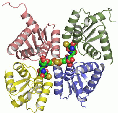
Chains, Units
Summary Information (see also Sequences/Alignments below) |
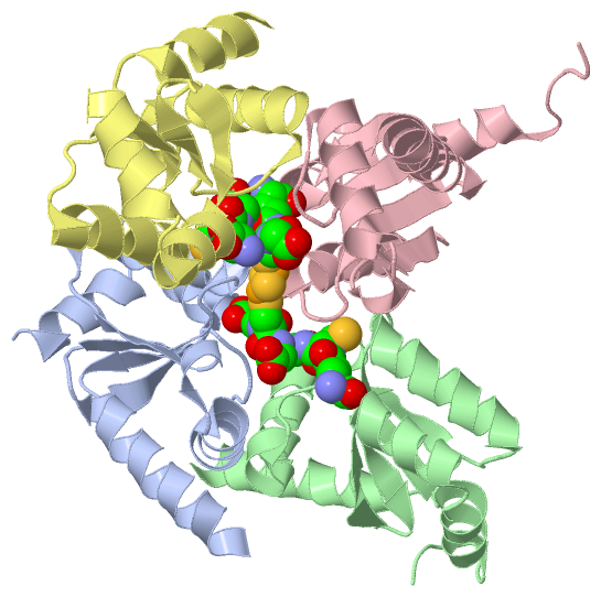
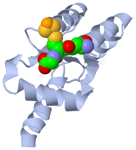
Ligands, Modified Residues, Ions (2, 5)| Asymmetric Unit (2, 5) Biological Unit 1 (2, 5) Biological Unit 2 (2, 2) Biological Unit 3 (1, 1) Biological Unit 4 (1, 1) Biological Unit 5 (1, 1) |
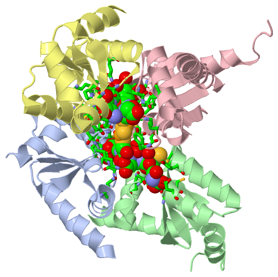
Sites (5, 5)
Asymmetric Unit (5, 5)
|
SS Bonds (0, 0)| (no "SS Bond" information available for 2E7P) |
Cis Peptide Bonds (4, 4)
Asymmetric Unit
|
||||||||||||||||||||
SAPs(SNPs)/Variants (0, 0)| (no "SAP(SNP)/Variant" information available for 2E7P) |
PROSITE Motifs (0, 0)| (no "PROSITE Motif" information available for 2E7P) |
Exons (0, 0)| (no "Exon" information available for 2E7P) |
Sequences/Alignments
Asymmetric UnitChain A from PDB Type:PROTEIN Length:107 aligned with Q5PSJ1_9ROSI | Q5PSJ1 from UniProtKB/TrEMBL Length:126 Alignment length:107 21 31 41 51 61 71 81 91 101 111 Q5PSJ1_9ROSI 12 SKQELDAALKKAKELASSAPVVVFSKTYCGYCNRVKQLLTQVGASYKVVELDELSDGSQLQSALAHWTGRGTVPNVFIGGKQIGGCDTVVEKHQRNELLPLLQDAAA 118 SCOP domains ----------------------------------------------------------------------------------------------------------- SCOP domains CATH domains ----------------------------------------------------------------------------------------------------------- CATH domains Pfam domains ----------------------------------------------------------------------------------------------------------- Pfam domains SAPs(SNPs) ----------------------------------------------------------------------------------------------------------- SAPs(SNPs) PROSITE ----------------------------------------------------------------------------------------------------------- PROSITE Transcript ----------------------------------------------------------------------------------------------------------- Transcript 2e7p A 2 SKQELDAALKKAKELASSAPVVVFSKTYCGYCNRVKQLLTQVGASYKVVELDELSDGSQLQSALAHWTGRGTVPNVFIGGKQIGGCDTVVEKHQRNELLPLLQDAAA 108 11 21 31 41 51 61 71 81 91 101 Chain B from PDB Type:PROTEIN Length:111 aligned with Q5PSJ1_9ROSI | Q5PSJ1 from UniProtKB/TrEMBL Length:126 Alignment length:111 22 32 42 52 62 72 82 92 102 112 122 Q5PSJ1_9ROSI 13 KQELDAALKKAKELASSAPVVVFSKTYCGYCNRVKQLLTQVGASYKVVELDELSDGSQLQSALAHWTGRGTVPNVFIGGKQIGGCDTVVEKHQRNELLPLLQDAAATAKTS 123 SCOP domains --------------------------------------------------------------------------------------------------------------- SCOP domains CATH domains --------------------------------------------------------------------------------------------------------------- CATH domains Pfam domains --------------------------------------------------------------------------------------------------------------- Pfam domains SAPs(SNPs) --------------------------------------------------------------------------------------------------------------- SAPs(SNPs) PROSITE --------------------------------------------------------------------------------------------------------------- PROSITE Transcript --------------------------------------------------------------------------------------------------------------- Transcript 2e7p B 3 KQELDAALKKAKELASSAPVVVFSKTYCGYCNRVKQLLTQVGASYKVVELDELSDGSQLQSALAHWTGRGTVPNVFIGGKQIGGCDTVVEKHQRNELLPLLQDAAATAKTS 113 12 22 32 42 52 62 72 82 92 102 112 Chain C from PDB Type:PROTEIN Length:115 aligned with Q5PSJ1_9ROSI | Q5PSJ1 from UniProtKB/TrEMBL Length:126 Alignment length:115 21 31 41 51 61 71 81 91 101 111 121 Q5PSJ1_9ROSI 12 SKQELDAALKKAKELASSAPVVVFSKTYCGYCNRVKQLLTQVGASYKVVELDELSDGSQLQSALAHWTGRGTVPNVFIGGKQIGGCDTVVEKHQRNELLPLLQDAAATAKTSAQL 126 SCOP domains ------------------------------------------------------------------------------------------------------------------- SCOP domains CATH domains ------------------------------------------------------------------------------------------------------------------- CATH domains Pfam domains ------------------------------------------------------------------------------------------------------------------- Pfam domains SAPs(SNPs) ------------------------------------------------------------------------------------------------------------------- SAPs(SNPs) PROSITE ------------------------------------------------------------------------------------------------------------------- PROSITE Transcript ------------------------------------------------------------------------------------------------------------------- Transcript 2e7p C 2 SKQELDAALKKAKELASSAPVVVFSKTYCGYCNRVKQLLTQVGASYKVVELDELSDGSQLQSALAHWTGRGTVPNVFIGGKQIGGCDTVVEKHQRNELLPLLQDAAATAKTSAQL 116 11 21 31 41 51 61 71 81 91 101 111 Chain D from PDB Type:PROTEIN Length:101 aligned with Q5PSJ1_9ROSI | Q5PSJ1 from UniProtKB/TrEMBL Length:126 Alignment length:101 26 36 46 56 66 76 86 96 106 116 Q5PSJ1_9ROSI 17 DAALKKAKELASSAPVVVFSKTYCGYCNRVKQLLTQVGASYKVVELDELSDGSQLQSALAHWTGRGTVPNVFIGGKQIGGCDTVVEKHQRNELLPLLQDAA 117 SCOP domains d2e7pd_ D: automated matches SCOP domains CATH domains ----------------------------------------------------------------------------------------------------- CATH domains Pfam domains ----------------------------------------------------------------------------------------------------- Pfam domains SAPs(SNPs) ----------------------------------------------------------------------------------------------------- SAPs(SNPs) PROSITE ----------------------------------------------------------------------------------------------------- PROSITE Transcript ----------------------------------------------------------------------------------------------------- Transcript 2e7p D 7 DAALKKAKELASSAPVVVFSKTYCGYCNRVKQLLTQVGASYKVVELDELSDGSQLQSALAHWTGRGTVPNVFIGGKQIGGCDTVVEKHQRNELLPLLQDAA 107 16 26 36 46 56 66 76 86 96 106
|
||||||||||||||||||||
SCOP Domains (1, 1)
Asymmetric Unit
 |
CATH Domains (0, 0)| (no "CATH Domain" information available for 2E7P) |
Pfam Domains (0, 0)| (no "Pfam Domain" information available for 2E7P) |
Gene Ontology (8, 8)|
Asymmetric Unit(hide GO term definitions) Chain A,B,C,D (Q5PSJ1_9ROSI | Q5PSJ1)
|
||||||||||||||||||||||||||||||||||||||||||||||||||||||||||||||||||
Interactive Views
|
|||||||||||||||||||||||||||||||||||||||||||||||||||||||||||||||||||||||||||||||||||||||||||||||||||||||||||||||||||||||||||||||||||||||||||||||||||||||||||||||||||||||||||||||||||||||||||||||||||||||||||||||||||||
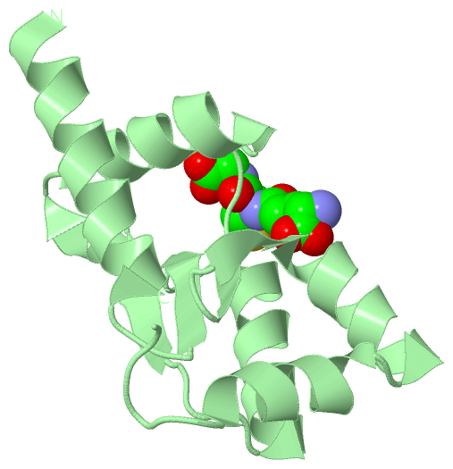
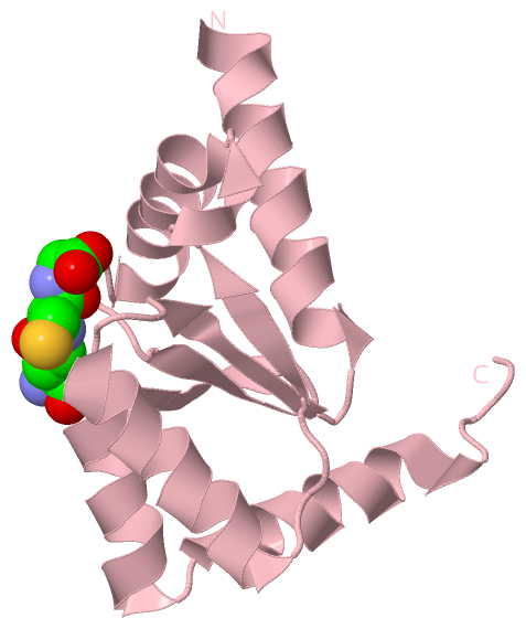
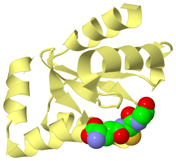
Still Images
|
||||||||||||||||
Databases
|
||||||||||||||||||||||||||||||||||||||||||||||||||||||||||||||||||||||||||||||||||||||||||||||||||||||||||||||||||||||||||||||||||||||||||||||||||||||||||||||||
Analysis Tools
|
|||||||||||||||||||||||||||||||||||||||||||||||||||||||||||||
Entries Sharing at Least One Protein Chain (UniProt ID)
Related Entries Specified in the PDB File
|
|