Show PDB file:   
         Plain Text   HTML   (compressed file size)
QuickSearch:   
by PDB,NDB,UniProt,PROSITE Code or Search Term(s)  
(-)Asymmetric Unit
(-)Asym. Unit - sites
(-)Biological Unit 1
(-)Biol. Unit 1 - sites
(-)Biological Unit 2
collapse expand < >
Image Asymmetric Unit
Asymmetric Unit  (Jmol Viewer)
Image Asym. Unit - sites
Asym. Unit - sites  (Jmol Viewer)
Image Biological Unit 1
Biological Unit 1  (Jmol Viewer)
Image Biol. Unit 1 - sites
Biol. Unit 1 - sites  (Jmol Viewer)
Image Biological Unit 2
Biological Unit 2  (Jmol Viewer)

(-) Description

Title :  THE CRYSTAL STRUCTURE OF EPOXIDE HYDROLASE B (RV1938) FROM MYCOBACTERIUM TUBERCULOSIS AT 2.1 ANGSTROM
 
Authors :  B. K. Biswal, Mycobacterium Tuberculosis Structural Proteomics (Xmtb)
Date :  27 Nov 06  (Deposition) - 04 Dec 07  (Release) - 13 Jul 11  (Revision)
Method :  X-RAY DIFFRACTION
Resolution :  2.10
Chains :  Asym. Unit :  A
Biol. Unit 1:  A  (2x)
Biol. Unit 2:  A  (1x)
Keywords :  Epoxide Hydrolase B, Mycobacterium Tuberculosis, Structural Genomics, Mycobacterium Tuberculosis Structural Proteomics Project, Xmtb, Hydrolase (Keyword Search: [Gene Ontology, PubMed, Web (Google))
 
Reference :  B. K. Biswal, C. Morisseau, G. Garen, M. M. Cherney, C. Garen, C. Niu, B. D. Hammock, M. N. James
The Molecular Structure Of Epoxide Hydrolase B From Mycobacterium Tuberculosis And Its Complex With A Urea-Base Inhibitor.
J. Mol. Biol. V. 381 897 2008
PubMed-ID: 18585390  |  Reference-DOI: 10.1016/J.JMB.2008.06.030

(-) Compounds

Molecule 1 - EPOXIDE HYDROLASE EPHB
    ChainsA
    EC Number3.3.2.3
    EngineeredYES
    Expression SystemESCHERICHIA COLI
    Expression System PlasmidPDEST17
    Expression System StrainBL21 DE3 (AI)
    Expression System Taxid562
    Expression System Vector TypePLASMID
    Organism ScientificMYCOBACTERIUM TUBERCULOSIS
    Organism Taxid83332
    StrainH37RV
    SynonymEPOXIDE HYDROLASE B, EPOXIDE HYDRATASE, EPOXIDE HYDROLASE

 Structural Features

(-) Chains, Units

  1
Asymmetric Unit A
Biological Unit 1 (2x)A
Biological Unit 2 (1x)A

Summary Information (see also Sequences/Alignments below)

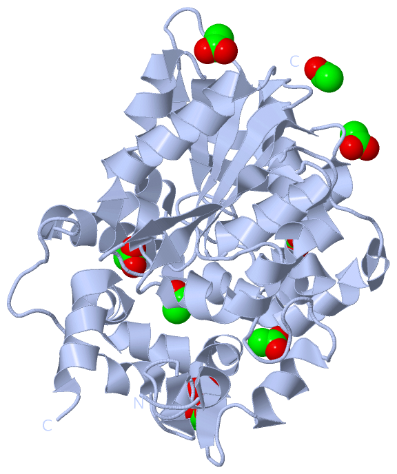
(-) Ligands, Modified Residues, Ions  (1, 9)

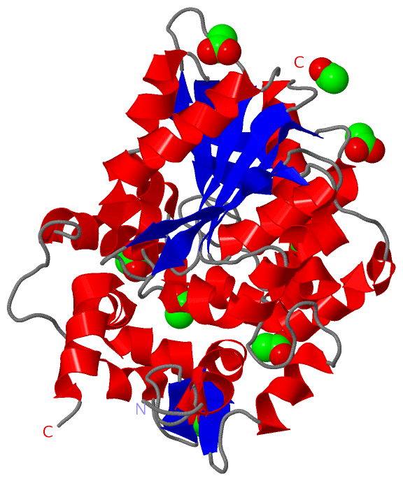
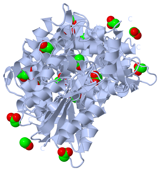
Asymmetric Unit (1, 9)
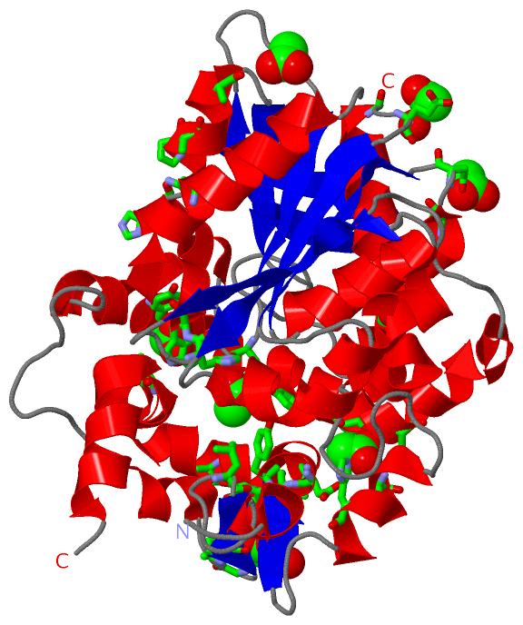
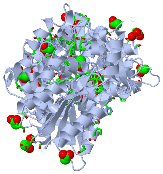
No.NameCountTypeFull Name
1ACT9Ligand/IonACETATE ION
Biological Unit 1 (1, 18)
No.NameCountTypeFull Name
1ACT18Ligand/IonACETATE ION
Biological Unit 2 (1, 9)
No.NameCountTypeFull Name
1ACT9Ligand/IonACETATE ION

(-) Sites  (9, 9)

Asymmetric Unit (9, 9)
No.NameEvidenceResiduesDescription
1AC1SOFTWAREVAL A:73 , THR A:176BINDING SITE FOR RESIDUE ACT A 2001
2AC2SOFTWARETYR A:167 , VAL A:170 , GLY A:173BINDING SITE FOR RESIDUE ACT A 2002
3AC3SOFTWARETHR A:190 , TYR A:256 , TRP A:334BINDING SITE FOR RESIDUE ACT A 2003
4AC4SOFTWARESER A:150 , HIS A:153 , ASP A:166 , TYR A:272 , HIS A:273 , ASP A:276BINDING SITE FOR RESIDUE ACT A 2004
5AC5SOFTWAREPHE A:36 , TYR A:164 , LEU A:226 , TYR A:272 , HIS A:333BINDING SITE FOR RESIDUE ACT A 2005
6AC6SOFTWAREALA A:53BINDING SITE FOR RESIDUE ACT A 2006
7AC7SOFTWAREMET A:228 , GLY A:231 , ALA A:232 , HOH A:2115BINDING SITE FOR RESIDUE ACT A 2007
8AC8SOFTWAREHIS A:45 , PRO A:48 , ALA A:49 , HIS A:117 , ASP A:119 , ASN A:344BINDING SITE FOR RESIDUE ACT A 2008
9AC9SOFTWAREGLY A:94 , GLU A:96 , HOH A:2132BINDING SITE FOR RESIDUE ACT A 2009

(-) SS Bonds  (0, 0)

(no "SS Bond" information available for 2E3J)

(-) Cis Peptide Bonds  (1, 1)

Asymmetric Unit
No.Residues
1Phe A:36 -Pro A:37

 Sequence-Structure Mapping

(-) SAPs(SNPs)/Variants  (0, 0)

(no "SAP(SNP)/Variant" information available for 2E3J)

(-) PROSITE Motifs  (0, 0)

(no "PROSITE Motif" information available for 2E3J)

(-) Exons   (0, 0)

(no "Exon" information available for 2E3J)

(-) Sequences/Alignments

Asymmetric Unit
   Reformat: Number of residues per line =  ('0' or empty: single-line sequence representation)
  Number of residues per labelling interval =   
  UniProt sequence: complete  aligned part    
   Show mapping: SCOP domains CATH domains Pfam domains Secondary structure (by author)
SAPs(SNPs) PROSITE motifs Exons
(details for a mapped element are shown in a popup box when the mouse pointer rests over it)
Chain A from PDB  Type:PROTEIN  Length:346
 aligned with EPHB_MYCTO | P95276 from UniProtKB/Swiss-Prot  Length:356

    Alignment length:353
                                    13        23        33        43        53        63        73        83        93       103       113       123       133       143       153       163       173       183       193       203       213       223       233       243       253       263       273       283       293       303       313       323       333       343       353   
           EPHB_MYCTO     4 VHRILNCRGTRIHAVADSPPDQQGPLVVLLHGFPESWYSWRHQIPALAGAGYRVVAIDQRGYGRSSKYRVQKAYRIKELVGDVVGVLDSYGAEQAFVVGHDWGAPVAWTFAWLHPDRCAGVVGISVPFAGRGVIGLPGSPFGERRPSDYHLELAGPGRVWYQDYFAVQDGIITEIEEDLRGWLLGLTYTVSGEGMMAATKAAVDAGVDLESMDPIDVIRAGPLCMAEGARLKDAFVYPETMPAWFTEADLDFYTGEFERSGFGGPLSFYHNIDNDWHDLADQQGKPLTPPALFIGGQYDVGTIWGAQAIERAHEVMPNYRGTHMIADVGHWIQQEAPEETNRLLLDFLGGLRP 356
               SCOP domains ----------------------------------------------------------------------------------------------------------------------------------------------------------------------------------------------------------------------------------------------------------------------------------------------------------------------------------------------------------------- SCOP domains
               CATH domains ----------------------------------------------------------------------------------------------------------------------------------------------------------------------------------------------------------------------------------------------------------------------------------------------------------------------------------------------------------------- CATH domains
               Pfam domains ----------------------------------------------------------------------------------------------------------------------------------------------------------------------------------------------------------------------------------------------------------------------------------------------------------------------------------------------------------------- Pfam domains
         Sec.struct. author .eeeeee..eeeeeeee........eeeee.....hhhhhh.hhhhhhhh..eeeee............hhhhhhhhhhhhhhhhhhhhh....eeeeee.hhhhhhhhhhhhh...eeeeeee....hhhhh...........hhhhhhhh.....eeehhhhhhhhhhhhhhhhhhhhhhhhhhhhhhhhhhhhhhhhh..-------..hhhhh......eee...hhhhhh..........hhhhhhhhhhhhhhhhhhhhhhhhhhhhhhhhhhhhhh.......eeeeee..hhhhhhhhhhhhhhhhhh..eeeeeee.....hhhhhhhhhhhhhhhhhhhh... Sec.struct. author
                 SAPs(SNPs) ----------------------------------------------------------------------------------------------------------------------------------------------------------------------------------------------------------------------------------------------------------------------------------------------------------------------------------------------------------------- SAPs(SNPs)
                    PROSITE ----------------------------------------------------------------------------------------------------------------------------------------------------------------------------------------------------------------------------------------------------------------------------------------------------------------------------------------------------------------- PROSITE
                 Transcript ----------------------------------------------------------------------------------------------------------------------------------------------------------------------------------------------------------------------------------------------------------------------------------------------------------------------------------------------------------------- Transcript
                 2e3j A   4 VHRILNCRGTRIHAVADSPPDQQGPLVVLLHGFPESWYSWRHQIPALAGAGYRVVAIDQRGYGRSSKYRVQKAYRIKELVGDVVGVLDSYGAEQAFVVGHDWGAPVAWTFAWLHPDRCAGVVGISVPFAGRGVIGLPGSPFGERRPSDYHLELAGPGRVWYQDYFAVQDGIITEIEEDLRGWLLGLTYTVSGEGMMAATKAAV-------SMDPIDVIRAGPLCMAEGARLKDAFVYPETMPAWFTEADLDFYTGEFERSGFGGPLSFYHNIDNDWHDLADQQGKPLTPPALFIGGQYDVGTIWGAQAIERAHEVMPNYRGTHMIADVGHWIQQEAPEETNRLLLDFLGGLRP 356
                                    13        23        33        43        53        63        73        83        93       103       113       123       133       143       153       163       173       183       193       203  |      -|      223       233       243       253       263       273       283       293       303       313       323       333       343       353   
                                                                                                                                                                                                                                    206     214                                                                                                                                              

   Legend:   → Mismatch (orange background)
  - → Gap (green background, '-', border residues have a numbering label)
    → Modified Residue (blue background, lower-case, 'x' indicates undefined single-letter code, labelled with number + name)
  x → Chemical Group (purple background, 'x', labelled with number + name, e.g. ACE or NH2)
  extra numbering lines below/above indicate numbering irregularities and modified residue names etc., number ends below/above '|'

 Classification and Annotation

(-) SCOP Domains  (0, 0)

(no "SCOP Domain" information available for 2E3J)

(-) CATH Domains  (0, 0)

(no "CATH Domain" information available for 2E3J)

(-) Pfam Domains  (0, 0)

(no "Pfam Domain" information available for 2E3J)

(-) Gene Ontology  (4, 4)

Asymmetric Unit(hide GO term definitions)
Chain A   (EPHB_MYCTO | P95276)
molecular function
    GO:0003824    catalytic activity    Catalysis of a biochemical reaction at physiological temperatures. In biologically catalyzed reactions, the reactants are known as substrates, and the catalysts are naturally occurring macromolecular substances known as enzymes. Enzymes possess specific binding sites for substrates, and are usually composed wholly or largely of protein, but RNA that has catalytic activity (ribozyme) is often also regarded as enzymatic.
    GO:0018742    epoxide hydrolase B activity    Catalysis of the hydrolysis of the ether in chloro- or hydroxyepoxypropane to produce chloropropane diol or glycerol. Acts on R enantiomers.
    GO:0016787    hydrolase activity    Catalysis of the hydrolysis of various bonds, e.g. C-O, C-N, C-C, phosphoric anhydride bonds, etc. Hydrolase is the systematic name for any enzyme of EC class 3.
    GO:0042803    protein homodimerization activity    Interacting selectively and non-covalently with an identical protein to form a homodimer.

 Visualization

(-) Interactive Views

Asymmetric Unit
  Complete Structure
    Jena3D(integrated viewing of ligand, site, SAP, PROSITE, SCOP information)
    WebMol | AstexViewer[tm]@PDBe
(Java Applets, require no local installation except for Java; loading may be slow)
    STRAP
(Java WebStart application, automatic local installation, requires Java; full application with system access!)
    RasMol
(require local installation)
    Molscript (VRML)
(requires installation of a VRML viewer; select preferred view via VRML and generate a mono or stereo PDF format file)
 
  Ligands, Modified Residues, Ions
    ACT  [ RasMol | Jena3D ]  +environment [ RasMol | Jena3D ]
 
  Sites
    AC1  [ RasMol ]  +environment [ RasMol ]
    AC2  [ RasMol ]  +environment [ RasMol ]
    AC3  [ RasMol ]  +environment [ RasMol ]
    AC4  [ RasMol ]  +environment [ RasMol ]
    AC5  [ RasMol ]  +environment [ RasMol ]
    AC6  [ RasMol ]  +environment [ RasMol ]
    AC7  [ RasMol ]  +environment [ RasMol ]
    AC8  [ RasMol ]  +environment [ RasMol ]
    AC9  [ RasMol ]  +environment [ RasMol ]
 
  Cis Peptide Bonds
    Phe A:36 - Pro A:37   [ RasMol ]  
 
Biological Units
  Complete Structure
    Biological Unit 1  [ Jena3D ]
    Biological Unit 2  [ Jena3D ]

(-) Still Images

Jmol
  protein: cartoon or spacefill or dots and stick; nucleic acid: cartoon and stick; ligands: spacefill; active site: stick
Molscript
  protein, nucleic acid: cartoon; ligands: spacefill; active site: ball and stick

 Databases and Analysis Tools

(-) Databases

Access by PDB/NDB ID
  2e3j
    Family and Domain InformationProDom | SYSTERS
    General Structural InformationGlycoscienceDB | MMDB | NDB | OCA | PDB | PDBe | PDBj | PDBsum | PDBWiki | PQS | PROTEOPEDIA
    Orientation in MembranesOPM
    Protein SurfaceSURFACE
    Secondary StructureDSSP (structure derived) | HSSP (homology derived)
    Structural GenomicsGeneCensus
    Structural NeighboursCE | VAST
    Structure ClassificationCATH | Dali | SCOP
    Validation and Original DataBMRB Data View | BMRB Restraints Grid | EDS | PROCHECK | RECOORD | WHAT_CHECK
 
Access by UniProt ID/Accession number
  EPHB_MYCTO | P95276
    Comparative Protein Structure ModelsModBase
    Genomic InformationEnsembl
    Protein-protein InteractionDIP
    Sequence, Family and Domain InformationInterPro | Pfam | SMART | UniProtKB/SwissProt
 
Access by Enzyme Classificator   (EC Number)
  3.3.2.3
    General Enzyme InformationBRENDA | EC-PDB | Enzyme | IntEnz
    PathwayKEGG | MetaCyc
 
Access by Disease Identifier   (MIM ID)
  (no 'MIM ID' available)
    Disease InformationOMIM
 
Access by GenAge ID
  (no 'GenAge ID' available)
    Age Related InformationGenAge

(-) Analysis Tools

Access by PDB/NDB ID
    Domain InformationXDom
    Interatomic Contacts of Structural UnitsCSU
    Ligand-protein ContactsLPC
    Protein CavitiescastP
    Sequence and Secondary StructurePDBCartoon
    Structure AlignmentSTRAP(Java WebStart application, automatic local installation, requires Java; full application with system access!)
    Structure and Sequence BrowserSTING
 
Access by UniProt ID/Accession number
  EPHB_MYCTO | P95276
    Protein Disorder PredictionDisEMBL | FoldIndex | GLOBPLOT (for more information see DisProt)

 Related Entries

(-) Entries Sharing at Least One Protein Chain (UniProt ID)

UniProtKB/Swiss-Prot
        EPHB_MYCTO | P952762zjf

(-) Related Entries Specified in the PDB File

(no "Related Entries Specified in the PDB File" available for 2E3J)