|
|
|
|
 Description
|
|
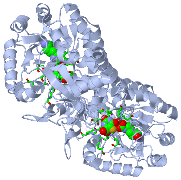
Compounds
|
||||||||||||||||||||||||||||||||||||||||||||||||||||||||
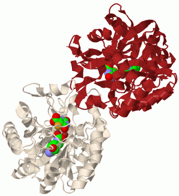
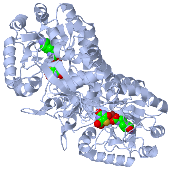
Chains, Units
Summary Information (see also Sequences/Alignments below) |
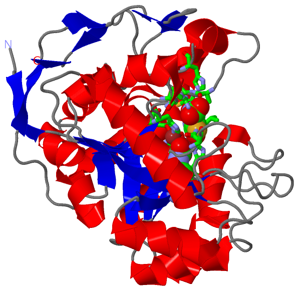
Ligands, Modified Residues, Ions (3, 4)| Asymmetric Unit (3, 4) Biological Unit 1 (2, 4) |
Sites (3, 3)
Asymmetric Unit (3, 3)
|
SS Bonds (0, 0)| (no "SS Bond" information available for 2E25) |
Cis Peptide Bonds (2, 2)
Asymmetric Unit
|
||||||||||||
SAPs(SNPs)/Variants (0, 0)| (no "SAP(SNP)/Variant" information available for 2E25) |
PROSITE Motifs (2, 2)
Asymmetric Unit (2, 2)
|
||||||||||||||||||||||||||||||||||||||||||||||||||||||||||||||||
Exons (0, 0)| (no "Exon" information available for 2E25) |
Sequences/Alignments
Asymmetric UnitChain A from PDB Type:PROTEIN Length:343 aligned with PYRC_ECOLI | P05020 from UniProtKB/Swiss-Prot Length:348 Alignment length:343 14 24 34 44 54 64 74 84 94 104 114 124 134 144 154 164 174 184 194 204 214 224 234 244 254 264 274 284 294 304 314 324 334 344 PYRC_ECOLI 5 SQVLKIRRPDDWHLHLRDGDMLKTVVPYTSEIYGRAIVMPNLAPPVTTVEAAVAYRQRILDAVPAGHDFTPLMTCYLTDSLDPNELERGFNEGVFTAAKLYPANATTNSSHGVTSIDAIMPVLERMEKIGMPLLVHGEVTHADIDIFDREARFIESVMEPLRQRLTALKVVFEHITTKDAADYVRDGNERLAATITPQHLMFNRNHMLVGGVRPHLYCLPILKRNIHQQALRELVASGFNRVFLGTDSAPHARHRKESSCGCAGCFNAPTALGSYATVFEEMNALQHFEAFCSVNGPQFYGLPVNDTFIELVREEQQVAESIALTDDTLVPFLAGETVRWSVK 347 SCOP domains d2e25a_ A: Dihydroorotase SCOP domains CATH domains 2e25A00 A:4-346 Metal-dependent hydrolases CATH domains Pfam domains ------------------------------------------------------------------------------------------------------------------------------------------------------------------------------------------------------------------------------------------------------------------------------------------------------------------------------------------------------- Pfam domains SAPs(SNPs) ------------------------------------------------------------------------------------------------------------------------------------------------------------------------------------------------------------------------------------------------------------------------------------------------------------------------------------------------------- SAPs(SNPs) PROSITE ----------DIHYDROOR---------------------------------------------------------------------------------------------------------------------------------------------------------------------------------------------------------------------------------DIHYDROOROTA--------------------------------------------------------------------------------------- PROSITE Transcript ------------------------------------------------------------------------------------------------------------------------------------------------------------------------------------------------------------------------------------------------------------------------------------------------------------------------------------------------------- Transcript 2e25 A 4 SQVLKIRRPDDWHLHLRDGDMLKTVVPYTSEIYGRAIVMPNLAPPVTTVEAAVAYRQRILDAVPAGHDFTPLMTCYLTDSLDPNELERGFNEGVFTAAkLYPANASTNSSHGVTSVDAIMPVLERMEKIGMPLLVHGEVTHADIDIFDREARFIESVMEPLRQRLTALKVVFEHITTKDAADYVRDGNERLAATITPQHLMFNRNHMLVGGVRPHLYCLPILKRNIHQQALRELVASGFNRVFLGTDSAPHARHRKESSCGCAGCFNAPTALGSYATVFEEMNALQHFEAFCSVNGPQFYGLPVNDTFIELVREEQQVAESIALTDDTLVPFLAGETVRWSVK 346 13 23 33 43 53 63 73 83 93 103 113 123 133 143 153 163 173 183 193 203 213 223 233 243 253 263 273 283 293 303 313 323 333 343 102-KCX
|
||||||||||||||||||||
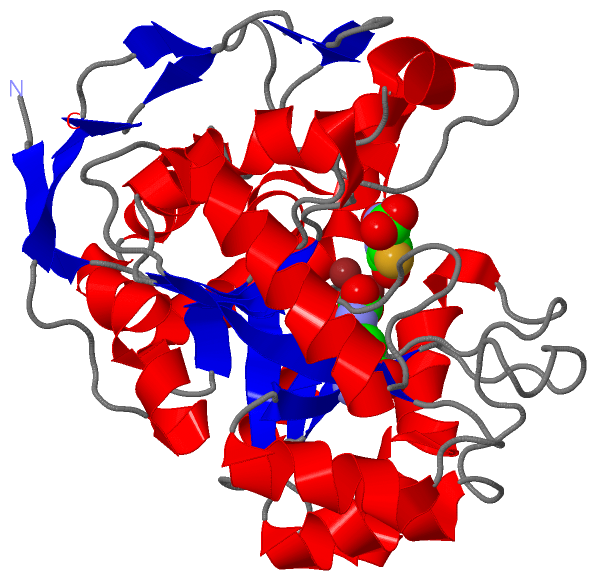
SCOP Domains (1, 1)
Asymmetric Unit

|
CATH Domains (1, 1)
Asymmetric Unit
|
Pfam Domains (0, 0)| (no "Pfam Domain" information available for 2E25) |
Gene Ontology (10, 10)|
Asymmetric Unit(hide GO term definitions) Chain A (PYRC_ECOLI | P05020)
|
||||||||||||||||||||||||||||||||||||||||||||||||||||||||||||||||||||||||||||||
Interactive Views
|
||||||||||||||||||||||||||||||||||||||||||||||||||||||||||||||||||||||||||||||||||||||||||||||||||||||||||||||||||||||||||||||||||||||||||||||||||||||||||||||||||||||||||||
Still Images
|
||||||||||||||||
Databases
|
||||||||||||||||||||||||||||||||||||||||||||||||||||||||||||||||||||||||||||||||||||||||||||||||||||||||||||||||||||||||||||||||||||||||||||||||||||||||||||||||
Analysis Tools
|
|||||||||||||||||||||||||||||||||||||||||||||||||||||||||||||
Entries Sharing at Least One Protein Chain (UniProt ID)
Related Entries Specified in the PDB File
|
|