|
|
|
|
 Description
|
|
Compounds
|
||||||||||||||||||||||||||||||||||||||||||||
Chains, Units
Summary Information (see also Sequences/Alignments below) |
Ligands, Modified Residues, Ions (0, 0)| (no "Ligand,Modified Residues,Ions" information available for 2DW3) |
Sites (0, 0)| (no "Site" information available for 2DW3) |
SS Bonds (0, 0)| (no "SS Bond" information available for 2DW3) |
Cis Peptide Bonds (0, 0)| (no "Cis Peptide Bond" information available for 2DW3) |
SAPs(SNPs)/Variants (0, 0)| (no "SAP(SNP)/Variant" information available for 2DW3) |
PROSITE Motifs (0, 0)| (no "PROSITE Motif" information available for 2DW3) |
Exons (0, 0)| (no "Exon" information available for 2DW3) |
Sequences/Alignments
NMR StructureChain A from PDB Type:PROTEIN Length:77 aligned with PUFX_RHOS4 | P13402 from UniProtKB/Swiss-Prot Length:82 Alignment length:79 11 21 31 41 51 61 71 PUFX_RHOS4 2 ADKTIFNDHLNTNPKTNLRLWVAFQMMKGAGWAGGVFFGTLLLIGFFRVVGRMLPIQENQAPAPNITGALETGIELIKH 80 SCOP domains ------------------------------------------------------------------------------- SCOP domains CATH domains 2dw3A01 A:1-55 ------------------------ CATH domains Pfam domains ------------------------------------------------------------------------------- Pfam domains SAPs(SNPs) ------------------------------------------------------------------------------- SAPs(SNPs) PROSITE ------------------------------------------------------------------------------- PROSITE Transcript ------------------------------------------------------------------------------- Transcript 2dw3 A 1 ADKTIFNDHLNTNPKTNLRLWVAFQMMKGAGWAGGVFFGTLLLIGFFRVVGRMLPIQENQAPAPNITGALEH--HHHHH 77 10 20 30 40 50 60 70 | | 72 73 Chain A from PDB Type:PROTEIN Length:77 aligned with Q7B2Z6_RHOSH | Q7B2Z6 from UniProtKB/TrEMBL Length:82 Alignment length:79 11 21 31 41 51 61 71 Q7B2Z6_RHOSH 2 ADKTIFNDHLNTNPKTNLRLWVAFQMMKGAGWAGGVFFGTLLLIGFFRVVGRMLPIQENQAPAPNITGALETGIELIKH 80 SCOP domains ------------------------------------------------------------------------------- SCOP domains CATH domains 2dw3A01 A:1-55 ------------------------ CATH domains Pfam domains ------------------------------------------------------------------------------- Pfam domains SAPs(SNPs) ------------------------------------------------------------------------------- SAPs(SNPs) PROSITE ------------------------------------------------------------------------------- PROSITE Transcript ------------------------------------------------------------------------------- Transcript 2dw3 A 1 ADKTIFNDHLNTNPKTNLRLWVAFQMMKGAGWAGGVFFGTLLLIGFFRVVGRMLPIQENQAPAPNITGALEH--HHHHH 77 10 20 30 40 50 60 70 | | 72 73
|
||||||||||||||||||||
SCOP Domains (0, 0)| (no "SCOP Domain" information available for 2DW3) |
CATH Domains (1, 1)
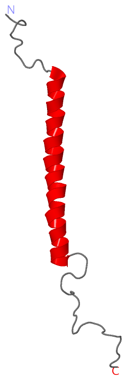
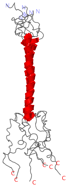
NMR Structure
 |
Pfam Domains (0, 0)| (no "Pfam Domain" information available for 2DW3) |
Gene Ontology (3, 3)|
NMR Structure(hide GO term definitions) Chain A (PUFX_RHOS4 | P13402)
|
||||||||||||||||||||||||||||||
Interactive Views
|
||||||||||||||||||||||||||||||||||||||||||||||||||||||||||||||||||||||||||||||||||||||||||||||||||||||||||||||||||||
Still Images
|
||||||||||||||||
Databases
|
||||||||||||||||||||||||||||||||||||||||||||||||||||||||||||||||||||||||||||||||||||||||||||||||||||||||||||||||||||||||||||||||||||||||||||||||||||||||||||||||||||||||||||||||||||||||||
Analysis Tools
|
||||||||||||||||||||||||||||||||||||||||||||||||||||||||||||||||||||||||
Entries Sharing at Least One Protein Chain (UniProt ID)
Related Entries Specified in the PDB File
|
|