|
|
|
|
 Description
|
|
Compounds
|
||||||||||||||||||||||||||||||||||||||||||||||||||||||||||||
Chains, Units
Summary Information (see also Sequences/Alignments below) |
Ligands, Modified Residues, Ions (1, 1)
NMR Structure (1, 1)
|
Sites (2, 2)
NMR Structure (2, 2)
|
SS Bonds (0, 0)| (no "SS Bond" information available for 2DVH) |
Cis Peptide Bonds (0, 0)| (no "Cis Peptide Bond" information available for 2DVH) |
SAPs(SNPs)/Variants (0, 0)| (no "SAP(SNP)/Variant" information available for 2DVH) |
PROSITE Motifs (0, 0)| (no "PROSITE Motif" information available for 2DVH) |
Exons (0, 0)| (no "Exon" information available for 2DVH) |
Sequences/Alignments
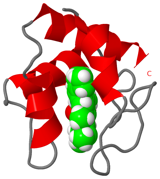
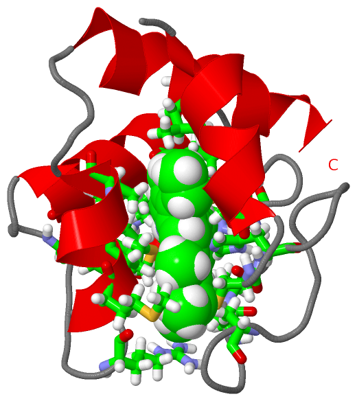
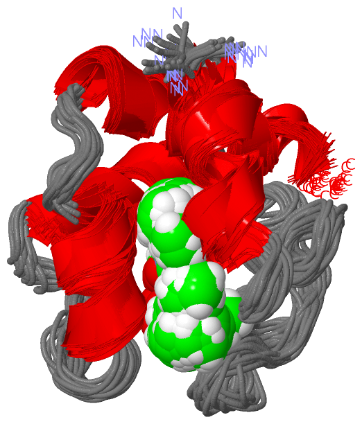
NMR StructureChain A from PDB Type:PROTEIN Length:79 aligned with CY553_DESVH | P04032 from UniProtKB/Swiss-Prot Length:103 Alignment length:79 34 44 54 64 74 84 94 CY553_DESVH 25 ADGAALYKSCIGCHGADGSKAAMGSAKPVKGQGAEELYKKMKGYADGSYGGERKAMMTNAVKKYSDEELKALADYMSKL 103 SCOP domains d2dvha_ A: Cytochrome c6 (synonym: cytochrome c553) SCOP domains CATH domains 2dvhA00 A:1-79 Cytochrome c CATH domains Pfam domains ------------------------------------------------------------------------------- Pfam domains SAPs(SNPs) ------------------------------------------------------------------------------- SAPs(SNPs) PROSITE ------------------------------------------------------------------------------- PROSITE Transcript ------------------------------------------------------------------------------- Transcript 2dvh A 1 ADGAALYKSCIGCHGADGSKAAMGSAKPVKGQGAEELYKKMKGYADGSYGGERKAMMTNAVKKASDEELKALADYMSKL 79 10 20 30 40 50 60 70
|
||||||||||||||||||||
SCOP Domains (1, 1)
NMR Structure
 |
CATH Domains (1, 1)
NMR Structure
|
Pfam Domains (0, 0)| (no "Pfam Domain" information available for 2DVH) |
Gene Ontology (5, 5)|
NMR Structure(hide GO term definitions) Chain A (CY553_DESVH | P04032)
|
||||||||||||||||||||||||||||||||||||||||||||||||
Interactive Views
|
|||||||||||||||||||||||||||||||||||||||||||||||||||||||||||||||||||||||||||||||||||||||||||||||||||||||||||||||||||||||||||||
Still Images
|
||||||||||||||||
Databases
|
||||||||||||||||||||||||||||||||||||||||||||||||||||||||||||||||||||||||||||||||||||||||||||||||||||||||||||||||||||||||||||||||||||||||||||||||||||||||||||||||
Analysis Tools
|
|||||||||||||||||||||||||||||||||||||||||||||||||||||||||||||
Entries Sharing at Least One Protein Chain (UniProt ID)
Related Entries Specified in the PDB File
|
|