|
|
|
|
 Description
|
|
Compounds
|
||||||||||||||||||||||||||||||||||||||||||||
Chains, Units
Summary Information (see also Sequences/Alignments below) |
Ligands, Modified Residues, Ions (2, 2)| Asymmetric Unit (2, 2) Biological Unit 1 (1, 1) Biological Unit 2 (1, 2) |
Sites (0, 0)| (no "Site" information available for 2DUY) |
SS Bonds (0, 0)| (no "SS Bond" information available for 2DUY) |
Cis Peptide Bonds (2, 2)
Asymmetric Unit
|
||||||||||||
SAPs(SNPs)/Variants (0, 0)| (no "SAP(SNP)/Variant" information available for 2DUY) |
PROSITE Motifs (0, 0)| (no "PROSITE Motif" information available for 2DUY) |
Exons (0, 0)| (no "Exon" information available for 2DUY) |
Sequences/Alignments
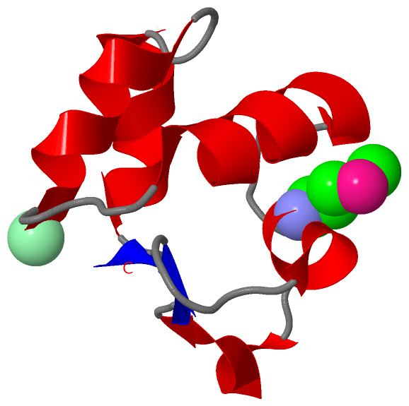
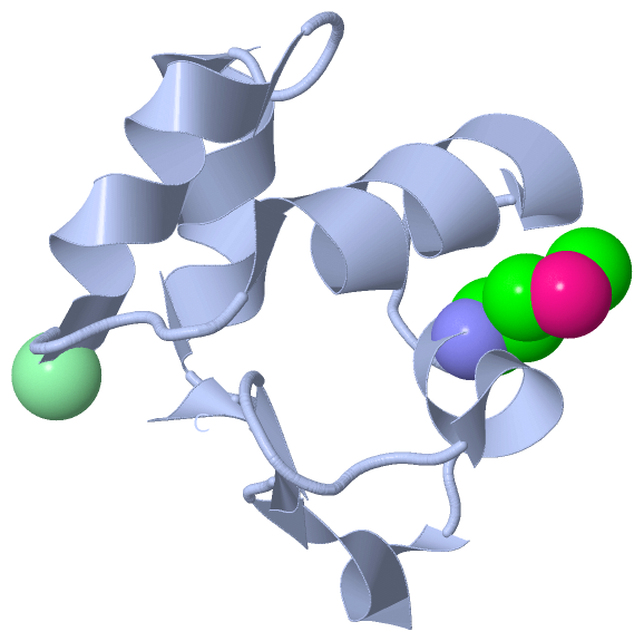
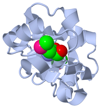
Asymmetric UnitChain A from PDB Type:PROTEIN Length:65 aligned with Q5SGW3_THET8 | Q5SGW3 from UniProtKB/TrEMBL Length:75 Alignment length:65 20 30 40 50 60 70 Q5SGW3_THET8 11 PLPQAQTPVSLNEASLEELMALPGIGPVLARRIVEGRPYARVEDLLKVKGIGPATLERLRPYLRP 75 SCOP domains d2duya1 A:11-75 Uncharacterized protein TTHA1967 SCOP domains CATH domains ----------------------------------------------------------------- CATH domains Pfam domains ----------------------------------------------------------------- Pfam domains SAPs(SNPs) ----------------------------------------------------------------- SAPs(SNPs) PROSITE ----------------------------------------------------------------- PROSITE Transcript ----------------------------------------------------------------- Transcript 2duy A 11 PLPQAQTPVSLNEASLEELmALPGIGPVLARRIVEGRPYARVEDLLKVKGIGPATLERLRPYLRP 75 20 30 40 50 60 70 30-MSE
|
||||||||||||||||||||
SCOP Domains (1, 1)
Asymmetric Unit
 |
CATH Domains (0, 0)| (no "CATH Domain" information available for 2DUY) |
Pfam Domains (0, 0)| (no "Pfam Domain" information available for 2DUY) |
Gene Ontology (2, 2)|
Asymmetric Unit(hide GO term definitions) Chain A (Q5SGW3_THET8 | Q5SGW3)
|
||||||||||||||||||||||||
Interactive Views
|
|||||||||||||||||||||||||||||||||||||||||||||||||||||||||||||||||||||||||||||||||||||||||||||||||||||||||||||||||||||||||||||||||||||||||||||||||||||||||||
Still Images
|
||||||||||||||||
Databases
|
||||||||||||||||||||||||||||||||||||||||||||||||||||||||||||||||||||||||||||||||||||||||||||||||||||||||||||||||||||||||||||||||||||||||||||||||||||||||||||||||
Analysis Tools
|
|||||||||||||||||||||||||||||||||||||||||||||||||||||||||||||
Entries Sharing at Least One Protein Chain (UniProt ID)
Related Entries Specified in the PDB File
|
|