Show PDB file:   
         Plain Text   HTML   (compressed file size)
QuickSearch:   
by PDB,NDB,UniProt,PROSITE Code or Search Term(s)  
(-)Asymmetric Unit
(-)Biological Unit 1
(-)Biological Unit 2
collapse expand < >
Image Asymmetric Unit
Asymmetric Unit  (Jmol Viewer)
Image Biological Unit 1
Biological Unit 1  (Jmol Viewer)
Image Biological Unit 2
Biological Unit 2  (Jmol Viewer)

(-) Description

Title :  CRYSTAL STRUCTURE OF MS0616
 
Authors :  T. Hosaka, A. Nishino, K. -T. Uchikubo, S. Kishishita, K. Murayama, M. Shirouzu, Riken Structural Genomics/Proteomics Initiative (Rsgi)
Date :  24 Jul 06  (Deposition) - 24 Jan 07  (Release) - 24 Feb 09  (Revision)
Method :  X-RAY DIFFRACTION
Resolution :  2.62
Chains :  Asym. Unit :  A,B
Biol. Unit 1:  A  (1x)
Biol. Unit 2:  B  (1x)
Keywords :  Hydrolase, Structural Genomics, Nppsfa, National Project On Protein Structural And Functional Analyses, Riken Structural Genomics/Proteomics Initiative, Rsgi (Keyword Search: [Gene Ontology, PubMed, Web (Google))
 
Reference :  T. Hosaka, A. Nishino, K. -T. Uchikubo, S. Kishishita, K. Murayama, M. Shirouzu, S. Yokoyama
Crystal Structure Of Ms0616
To Be Published
PubMed: search
(for further references see the PDB file header)

(-) Compounds

Molecule 1 - MS0616
    ChainsA, B
    EC Number3.6.1.52, 3.6.1.-
    EngineeredYES
    Expression System PlasmidPX051128-08
    Expression System Vector TypePLASMID
    Organism CommonHOUSE MOUSE
    Organism ScientificMUS MUSCULUS
    Organism Taxid10090
    Other DetailsCELL-FREE PROTEIN SYNTHESIS
    SynonymDIPHOSPHOINOSITOL POLYPHOSPHATE PHOSPHOHYDROLASE 2, DIPP-2, DIADENOSINE 5',5'''-P1,P6-HEXAPHOSPHATE HYDROLASE 2, NUCLEOSIDE DIPHOSPHATE-LINKED MOIETY X MOTIF 4, NUDIX MOTIF 4

 Structural Features

(-) Chains, Units

  12
Asymmetric Unit AB
Biological Unit 1 (1x)A 
Biological Unit 2 (1x) B

Summary Information (see also Sequences/Alignments below)

(-) Ligands, Modified Residues, Ions  (0, 0)

(no "Ligand,Modified Residues,Ions" information available for 2DUK)

(-) Sites  (0, 0)

(no "Site" information available for 2DUK)

(-) SS Bonds  (1, 1)

Asymmetric Unit
No.Residues
1A:123 -B:123

(-) Cis Peptide Bonds  (0, 0)

(no "Cis Peptide Bond" information available for 2DUK)

 Sequence-Structure Mapping

(-) SAPs(SNPs)/Variants  (0, 0)

(no "SAP(SNP)/Variant" information available for 2DUK)

(-) PROSITE Motifs  (2, 4)

Asymmetric Unit (2, 4)
 PROSITEUniProtKBPDB
No.IDACDescriptionIDLocationCountLocation
1NUDIXPS51462 Nudix hydrolase domain profile.NUDT4_MOUSE17-143
 
  2A:10-136
B:10-136
2NUDIX_BOXPS00893 Nudix box signature.NUDT4_MOUSE50-71
 
  2A:43-64
B:43-64
Biological Unit 1 (2, 2)
 PROSITEUniProtKBPDB
No.IDACDescriptionIDLocationCountLocation
1NUDIXPS51462 Nudix hydrolase domain profile.NUDT4_MOUSE17-143
 
  1A:10-136
-
2NUDIX_BOXPS00893 Nudix box signature.NUDT4_MOUSE50-71
 
  1A:43-64
-
Biological Unit 2 (2, 2)
 PROSITEUniProtKBPDB
No.IDACDescriptionIDLocationCountLocation
1NUDIXPS51462 Nudix hydrolase domain profile.NUDT4_MOUSE17-143
 
  1-
B:10-136
2NUDIX_BOXPS00893 Nudix box signature.NUDT4_MOUSE50-71
 
  1-
B:43-64

(-) Exons   (0, 0)

(no "Exon" information available for 2DUK)

(-) Sequences/Alignments

Asymmetric Unit
   Reformat: Number of residues per line =  ('0' or empty: single-line sequence representation)
  Number of residues per labelling interval =   
  UniProt sequence: complete  aligned part    
   Show mapping: SCOP domains CATH domains Pfam domains Secondary structure (by author)
SAPs(SNPs) PROSITE motifs Exons
(details for a mapped element are shown in a popup box when the mouse pointer rests over it)
Chain A from PDB  Type:PROTEIN  Length:138
 aligned with NUDT4_MOUSE | Q8R2U6 from UniProtKB/Swiss-Prot  Length:179

    Alignment length:138
                                    17        27        37        47        57        67        77        87        97       107       117       127       137        
          NUDT4_MOUSE     8 TRTYDREGFKKRAACLCFRSEQEDEVLLVSSSRYPDQWIVPGGGMEPEEEPGGAAVREVYEEAGVKGKLGRLLGIFENQDRKHRTYVYVLTVTEILEDWEDSVNIGRKREWFKVEDAIKVLQCHKPVHAEYLEKLKLG 145
               SCOP domains d2duka_ A: automated matches                                                                                                               SCOP domains
               CATH domains ------------------------------------------------------------------------------------------------------------------------------------------ CATH domains
               Pfam domains ------------------------------------------------------------------------------------------------------------------------------------------ Pfam domains
         Sec.struct. author .........eeeeeeeee......eeeeee.......ee..eee.....hhhhhhhhhhhhhh.eeeeeeeeeeeeee....eeeeeeeeeeeee.....hhhhhh..eeeeehhhhhhhhhh.hhhhhhhhh..... Sec.struct. author
                 SAPs(SNPs) ------------------------------------------------------------------------------------------------------------------------------------------ SAPs(SNPs)
                PROSITE (1) ---------NUDIX  PDB: A:10-136 UniProt: 17-143                                                                                           -- PROSITE (1)
                PROSITE (2) ------------------------------------------NUDIX_BOX  PDB: A:43-6-------------------------------------------------------------------------- PROSITE (2)
                 Transcript ------------------------------------------------------------------------------------------------------------------------------------------ Transcript
                 2duk A   1 TRTYDREGFKKRAACLCFRSEQEDEVLLVSSSRYPDQWIVPGGGMEPEEEPGGAAVREVYEEAGVKGKLGRLLGIFENQDRKHRTYVYVLTVTEILEDWEDSVNIGRKREWFKVEDAIKVLQCHKPVHAEYLEKLKLG 138
                                    10        20        30        40        50        60        70        80        90       100       110       120       130        

Chain B from PDB  Type:PROTEIN  Length:132
 aligned with NUDT4_MOUSE | Q8R2U6 from UniProtKB/Swiss-Prot  Length:179

    Alignment length:138
                                    17        27        37        47        57        67        77        87        97       107       117       127       137        
          NUDT4_MOUSE     8 TRTYDREGFKKRAACLCFRSEQEDEVLLVSSSRYPDQWIVPGGGMEPEEEPGGAAVREVYEEAGVKGKLGRLLGIFENQDRKHRTYVYVLTVTEILEDWEDSVNIGRKREWFKVEDAIKVLQCHKPVHAEYLEKLKLG 145
               SCOP domains d2dukb_ B: automated matches                                                                                                               SCOP domains
               CATH domains ------------------------------------------------------------------------------------------------------------------------------------------ CATH domains
               Pfam domains ------------------------------------------------------------------------------------------------------------------------------------------ Pfam domains
         Sec.struct. author .........eeeeeeeee......eeeeee.......ee..eee.....hhhhhhhhhhhhh..eeeeeeeeeeeeee....eeeeeeeeeeeee....------...eeeeehhhhhhhhh....hhhhhhhhhh.. Sec.struct. author
                 SAPs(SNPs) ------------------------------------------------------------------------------------------------------------------------------------------ SAPs(SNPs)
                PROSITE (1) ---------NUDIX  PDB: B:10-136 UniProt: 17-143                                                                                           -- PROSITE (1)
                PROSITE (2) ------------------------------------------NUDIX_BOX  PDB: B:43-6-------------------------------------------------------------------------- PROSITE (2)
                 Transcript ------------------------------------------------------------------------------------------------------------------------------------------ Transcript
                 2duk B   1 TRTYDREGFKKRAACLCFRSEQEDEVLLVSSSRYPDQWIVPGGGMEPEEEPGGAAVREVYEEAGVKGKLGRLLGIFENQDRKHRTYVYVLTVTEILEDW------GRKREWFKVEDAIKVLQCHKPVHAEYLEKLKLG 138
                                    10        20        30        40        50        60        70        80        90        |-     | 110       120       130        
                                                                                                                             99    106                                

   Legend:   → Mismatch (orange background)
  - → Gap (green background, '-', border residues have a numbering label)
    → Modified Residue (blue background, lower-case, 'x' indicates undefined single-letter code, labelled with number + name)
  x → Chemical Group (purple background, 'x', labelled with number + name, e.g. ACE or NH2)
  extra numbering lines below/above indicate numbering irregularities and modified residue names etc., number ends below/above '|'

 Classification and Annotation

(-) SCOP Domains  (1, 2)

Asymmetric Unit

(-) CATH Domains  (0, 0)

(no "CATH Domain" information available for 2DUK)

(-) Pfam Domains  (0, 0)

(no "Pfam Domain" information available for 2DUK)

(-) Gene Ontology  (17, 19)

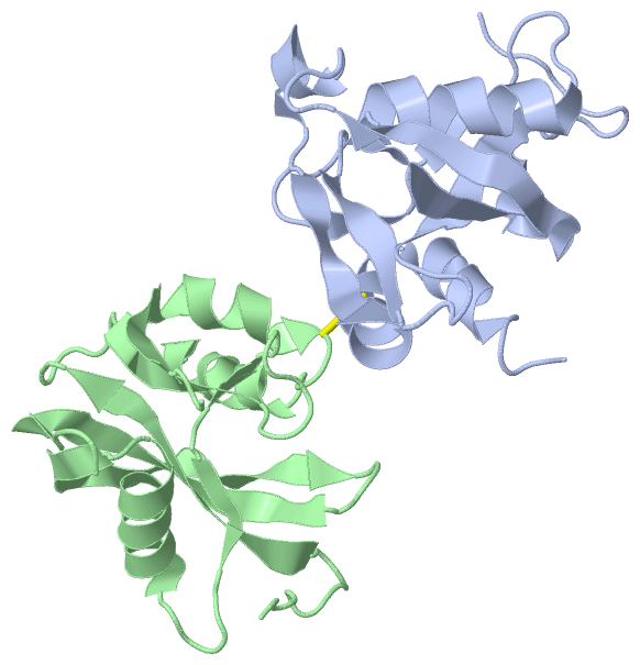
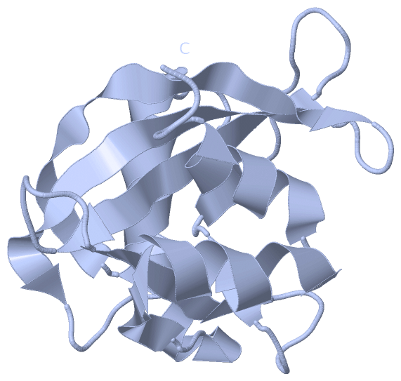
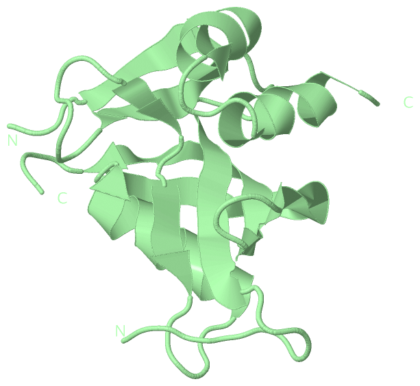
Asymmetric Unit(hide GO term definitions)
Chain A,B   (NUDT4_MOUSE | Q8R2U6)
molecular function
    GO:0003723    RNA binding    Interacting selectively and non-covalently with an RNA molecule or a portion thereof.
    GO:0008486    diphosphoinositol-polyphosphate diphosphatase activity    Catalysis of the reaction: diphospho-myo-inositol polyphosphate + H2O = myo-inositol polyphosphate + phosphate.
    GO:0016787    hydrolase activity    Catalysis of the hydrolysis of various bonds, e.g. C-O, C-N, C-C, phosphoric anhydride bonds, etc. Hydrolase is the systematic name for any enzyme of EC class 3.
    GO:0052841    inositol bisdiphosphate tetrakisphosphate diphosphatase activity    Catalysis of the reaction: inositol bisdiphosphate tetrakisphosphate + H2O = inositol diphosphate pentakisphosphate + phosphate.
    GO:0052842    inositol diphosphate pentakisphosphate diphosphatase activity    Catalysis of the reaction: inositol diphosphate pentakisphosphate + H2O = inositol hexakisphosphate + phosphate.
    GO:0052840    inositol diphosphate tetrakisphosphate diphosphatase activity    Catalysis of the reaction: inositol diphosphate tetrakisphosphate + H2O = inositol 1,3,4,5,6-pentakisphosphate + phosphate.
    GO:0052846    inositol-1,5-bisdiphosphate-2,3,4,6-tetrakisphosphate 1-diphosphatase activity    Catalysis of the reaction: 1,5-bisdiphosphoinositol-1D-myo-inositol 2,3,4,6-tetrakisphosphate + H2O = 1-diphospho-1D-myo-inositol 1,2,3,4,6-pentakisphosphate + phosphate + H+.
    GO:0052847    inositol-1,5-bisdiphosphate-2,3,4,6-tetrakisphosphate 5-diphosphatase activity    Catalysis of the reaction: 1,5-bisdiphosphoinositol-1D-myo-inositol 2,3,4,6-tetrakisphosphate + H2O = 1-diphospho-1D-myo-inositol 2,3,4,5,6-pentakisphosphate + phosphate + H+.
    GO:0052843    inositol-1-diphosphate-2,3,4,5,6-pentakisphosphate diphosphatase activity    Catalysis of the reaction: inositol 1-diphosphate 2,3,4,5,6-pentakisphosphate + H2O = inositol 1,2,3,4,5,6-hexakisphosphate + phosphate + 2 H+.
    GO:0052848    inositol-3,5-bisdiphosphate-2,3,4,6-tetrakisphosphate 5-diphosphatase activity    Catalysis of the reaction: 3,5-bisdiphosphoinositol-1D-myo-inositol 2,3,4,6-tetrakisphosphate + H2O = 3-diphospho-1D-myo-inositol 1,2,4,5,6-pentakisphosphate + phosphate + H+.
    GO:0052844    inositol-3-diphosphate-1,2,4,5,6-pentakisphosphate diphosphatase activity    Catalysis of the reaction: inositol 3-diphosphate 1,2,4,5,6-pentakisphosphate + H2O = inositol 1,2,3,4,5,6-hexakisphosphate + phosphate + 2 H+.
    GO:0052845    inositol-5-diphosphate-1,2,3,4,6-pentakisphosphate diphosphatase activity    Catalysis of the reaction: inositol 5-diphosphate 1,2,3,4,6-pentakisphosphate + H2O = inositol 1,2,3,4,5,6-hexakisphosphate + phosphate + 2 H+.
    GO:0050072    m7G(5')pppN diphosphatase activity    Catalysis of the reaction: 7-methylguanosine 5'-triphospho-5'-polynucleotide + H2O = 7-methylguanosine 5'-phosphate + polynucleotide.
    GO:0046872    metal ion binding    Interacting selectively and non-covalently with any metal ion.
    GO:0030515    snoRNA binding    Interacting selectively and non-covalently with small nucleolar RNA.
cellular component
    GO:0005575    cellular_component    The part of a cell, extracellular environment or virus in which a gene product is located. A gene product may be located in one or more parts of a cell and its location may be as specific as a particular macromolecular complex, that is, a stable, persistent association of macromolecules that function together.
    GO:0005737    cytoplasm    All of the contents of a cell excluding the plasma membrane and nucleus, but including other subcellular structures.

 Visualization

(-) Interactive Views

Asymmetric Unit
  Complete Structure
    Jena3D(integrated viewing of ligand, site, SAP, PROSITE, SCOP information)
    WebMol | AstexViewer[tm]@PDBe
(Java Applets, require no local installation except for Java; loading may be slow)
    STRAP
(Java WebStart application, automatic local installation, requires Java; full application with system access!)
    RasMol
(require local installation)
    Molscript (VRML)
(requires installation of a VRML viewer; select preferred view via VRML and generate a mono or stereo PDF format file)
 
  Ligands, Modified Residues, Ions
(no "Ligands, Modified Residues, Ions" information available for 2duk)
 
  Sites
(no "Sites" information available for 2duk)
 
  Cis Peptide Bonds
(no "Cis Peptide Bonds" information available for 2duk)
 
Biological Units
  Complete Structure
    Biological Unit 1  [ Jena3D ]
    Biological Unit 2  [ Jena3D ]

(-) Still Images

Jmol
  protein: cartoon or spacefill or dots and stick; nucleic acid: cartoon and stick; ligands: spacefill; active site: stick
Molscript
  protein, nucleic acid: cartoon; ligands: spacefill; active site: ball and stick

 Databases and Analysis Tools

(-) Databases

Access by PDB/NDB ID
  2duk
    Family and Domain InformationProDom | SYSTERS
    General Structural InformationGlycoscienceDB | MMDB | NDB | OCA | PDB | PDBe | PDBj | PDBsum | PDBWiki | PQS | PROTEOPEDIA
    Orientation in MembranesOPM
    Protein SurfaceSURFACE
    Secondary StructureDSSP (structure derived) | HSSP (homology derived)
    Structural GenomicsGeneCensus
    Structural NeighboursCE | VAST
    Structure ClassificationCATH | Dali | SCOP
    Validation and Original DataBMRB Data View | BMRB Restraints Grid | EDS | PROCHECK | RECOORD | WHAT_CHECK
 
Access by UniProt ID/Accession number
  NUDT4_MOUSE | Q8R2U6
    Comparative Protein Structure ModelsModBase
    Genomic InformationEnsembl
    Protein-protein InteractionDIP
    Sequence, Family and Domain InformationInterPro | Pfam | SMART | UniProtKB/SwissProt
 
Access by Enzyme Classificator   (EC Number)
  3.6.1.-
    General Enzyme InformationBRENDA | EC-PDB | Enzyme | IntEnz
    PathwayKEGG | MetaCyc
  3.6.1.52
    General Enzyme InformationBRENDA | EC-PDB | Enzyme | IntEnz
    PathwayKEGG | MetaCyc
 
Access by Disease Identifier   (MIM ID)
  (no 'MIM ID' available)
    Disease InformationOMIM
 
Access by GenAge ID
  (no 'GenAge ID' available)
    Age Related InformationGenAge

(-) Analysis Tools

Access by PDB/NDB ID
    Domain InformationXDom
    Interatomic Contacts of Structural UnitsCSU
    Ligand-protein ContactsLPC
    Protein CavitiescastP
    Sequence and Secondary StructurePDBCartoon
    Structure AlignmentSTRAP(Java WebStart application, automatic local installation, requires Java; full application with system access!)
    Structure and Sequence BrowserSTING
 
Access by UniProt ID/Accession number
  NUDT4_MOUSE | Q8R2U6
    Protein Disorder PredictionDisEMBL | FoldIndex | GLOBPLOT (for more information see DisProt)

 Related Entries

(-) Entries Sharing at Least One Protein Chain (UniProt ID)

(no "Entries Sharing at Least One Protein Chain" available for 2DUK)

(-) Related Entries Specified in the PDB File

(no "Related Entries Specified in the PDB File" available for 2DUK)