|
|
|
|
 Description
|
|
Compounds
|
||||||||||||||||||||||||||||||||||||||||||||||||||||
Chains, Units
Summary Information (see also Sequences/Alignments below) |
Ligands, Modified Residues, Ions (0, 0)| (no "Ligand,Modified Residues,Ions" information available for 2DJK) |
Sites (0, 0)| (no "Site" information available for 2DJK) |
SS Bonds (0, 0)| (no "SS Bond" information available for 2DJK) |
Cis Peptide Bonds (1, 10)
NMR Structure
|
||||||||||
SAPs(SNPs)/Variants (0, 0)| (no "SAP(SNP)/Variant" information available for 2DJK) |
PROSITE Motifs (1, 1)
NMR Structure (1, 1)
|
||||||||||||||||||||||||
Exons (0, 0)| (no "Exon" information available for 2DJK) |
Sequences/Alignments
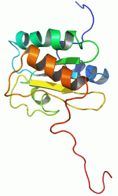
NMR StructureChain A from PDB Type:PROTEIN Length:133 aligned with PDI_HUMIN | P55059 from UniProtKB/Swiss-Prot Length:505 Alignment length:181 184 194 204 214 224 234 244 254 264 274 284 294 304 314 324 334 344 354 PDI_HUMIN 175 YPFGSSSDAALAEAEGVKAPAIVLYKDFDEGKAVFSEKFEVEAIEKFAKTGATPLIGEIGPETYSDYMSAGIPLAYIFAETAEERKELSDKLKPIAEAQRGVINFGTIDAKAFGAHAGNLNLKTDKFPAFAIQEVAKNQKFPFDQEKEITFEAIKAFVDDFVAGKIEPSIKSEPIPEKQEG 355 SCOP domains d2djk a1 A:1-133 Protein disulfide isomerase, PDI SCOP domains CATH domains ------------------------------------------------------------------------------------------------------------------------------------------------------------------------------------- CATH domains Pfam domains ------------------------------------------------------------------------------------------------------------------------------------------------------------------------------------- Pfam domains SAPs(SNPs) ------------------------------------------------------------------------------------------------------------------------------------------------------------------------------------- SAPs(SNPs) PROSITE ----------------------------------------------------------------------------------------------------------------------------------------------------------------THIOREDOXIN_2 PROSITE Transcript ------------------------------------------------------------------------------------------------------------------------------------------------------------------------------------- Transcript 2djk A 1 GPLGS------------------------------------------------PLIGEIGPETYSDYMSAGIPLAYIFAETAEERKELSDKLKPIAEAQRGVINFGTIDAKAFGAHAGNLNLKTDKFPAFAIQEVAKNQKFPFDQEKEITFEAIKAFVDDFVAGKIEPSIKSEPIPEKQEG 133 | - - - - - | 12 22 32 42 52 62 72 82 92 102 112 122 132 5 6
|
||||||||||||||||||||
SCOP Domains (1, 1)
NMR Structure
 |
CATH Domains (0, 0)| (no "CATH Domain" information available for 2DJK) |
Pfam Domains (0, 0)| (no "Pfam Domain" information available for 2DJK) |
Gene Ontology (5, 5)|
NMR Structure(hide GO term definitions) Chain A (PDI_HUMIN | P55059)
|
||||||||||||||||||||||||||||||||||||||||||||||||
Interactive Views
|
|||||||||||||||||||||||||||||||||||||||||||||||||||||||||||||||||||||||||||||||||||||||||||||||||||||||||||||||||||||
Still Images
|
||||||||||||||||
Databases
|
||||||||||||||||||||||||||||||||||||||||||||||||||||||||||||||||||||||||||||||||||||||||||||||||||||||||||||||||||||||||||||||||||||||||||||||||||||||||||||||||
Analysis Tools
|
|||||||||||||||||||||||||||||||||||||||||||||||||||||||||||||
Entries Sharing at Least One Protein Chain (UniProt ID)
Related Entries Specified in the PDB File
|
|