|
|
|
|
 Description
|
|
Compounds
|
||||||||||||||||||||||||||||||||||||||||||||||||||||||||
Chains, Units
Summary Information (see also Sequences/Alignments below) |
Ligands, Modified Residues, Ions (0, 0)| (no "Ligand,Modified Residues,Ions" information available for 2DJ1) |
Sites (0, 0)| (no "Site" information available for 2DJ1) |
SS Bonds (0, 0)| (no "SS Bond" information available for 2DJ1) |
Cis Peptide Bonds (2, 40)
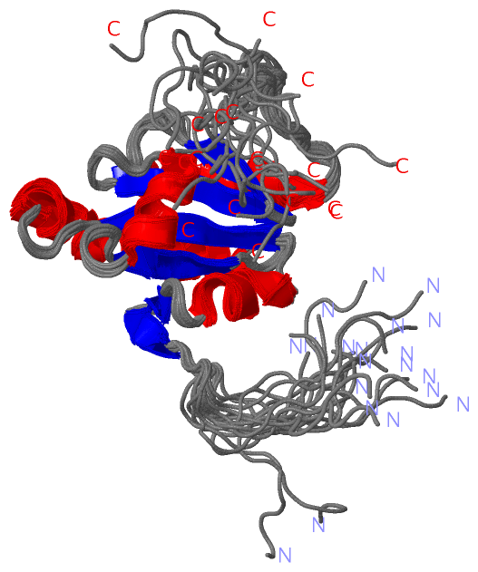
NMR Structure
|
|||||||||||||||
SAPs(SNPs)/Variants (0, 0)| (no "SAP(SNP)/Variant" information available for 2DJ1) |
PROSITE Motifs (2, 2)
NMR Structure (2, 2)
|
||||||||||||||||||||||||||||||||
Exons (0, 0)| (no "Exon" information available for 2DJ1) |
Sequences/Alignments
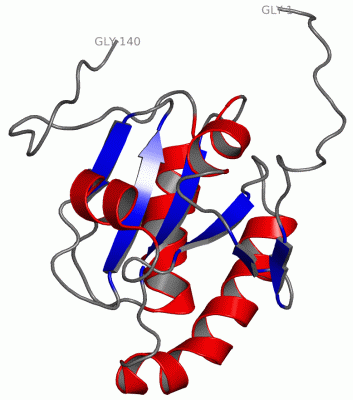
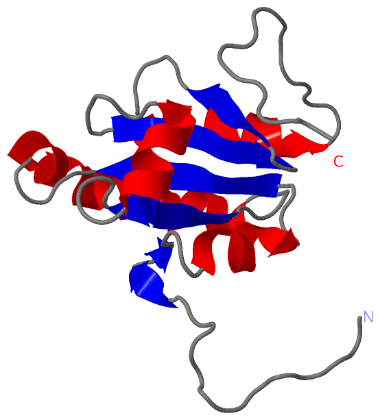
NMR StructureChain A from PDB Type:PROTEIN Length:140 aligned with PDIA4_MOUSE | P08003 from UniProtKB/Swiss-Prot Length:638 Alignment length:260 31 41 51 61 71 81 91 101 111 121 131 141 151 161 171 181 191 201 211 221 231 241 251 261 271 281 PDIA4_MOUSE 22 ASAGDAHEDTSDTENATEEEEEEDDDDLEVKEENGVWVLNDGNFDNFVADKDTVLLEFYAPWCGHCKQFAPEYEKIASTLKDNDPPIAVAKIDATSASMLASKFDVSGYPTIKILKKGQAVDYDGSRTQEEIVAKVREVSQPDWTPPPEVTLSLTKDNFDDVVNNADIILVEFYAPWCGHCKKLAPEYEKAAKELSKRSPPIPLAKVDATEQTDLAKRFDVSGYPTLKIFRKGRPFDYNGPREKYGIVDYMIEQSGPPSK 281 SCOP domains d2dj1a _ A: automated matches SCOP domains CATH domains -------------------------------------------------------------------------------------------------------------------------------------------------------------------------------------------------------------------------------------------------------------------- CATH domains Pfam domains -------------------------------------------------------------------------------------------------------------------------------------------------------------------------------------------------------------------------------------------------------------------- Pfam domains SAPs(SNPs) -------------------------------------------------------------------------------------------------------------------------------------------------------------------------------------------------------------------------------------------------------------------- SAPs(SNPs) PROSITE (1) ------------------------------------------------------THIOREDOXIN_1 ------------------------------------------------------------------------------------------------THIOREDOXIN_1 ------------------------------------------------------------------------ PROSITE (1) PROSITE (2) THIOREDOXIN_2 PDB: - UniProt: 16-160 -THIOREDOXIN_2 PDB: A:124-139 UniProt: 162-294 PROSITE (2) Transcript -------------------------------------------------------------------------------------------------------------------------------------------------------------------------------------------------------------------------------------------------------------------- Transcript 2dj1 A 1 GSSGSS-----------------GDDDLEVKEENGVWVLNDGNFDNFVADKDTVLLEFYAPWCGHCKQFAPEYEKIASTLKDNDPPIAVAKIDATSASMLASKFDVSGYPTIKILKKGQAVDYDGSRTQEEIVAKVREVSQPDWTPPPEVT-------------------------------------------------------------------------------------------------------SGPSSG 140 | - - | 13 23 33 43 53 63 73 83 93 103 113 123 133| - - - - - - - - - - | 140 6 7 134 135
|
||||||||||||||||||||
SCOP Domains (1, 1)
NMR Structure
 |
CATH Domains (0, 0)| (no "CATH Domain" information available for 2DJ1) |
Pfam Domains (0, 0)| (no "Pfam Domain" information available for 2DJ1) |
Gene Ontology (11, 11)|
NMR Structure(hide GO term definitions) Chain A (PDIA4_MOUSE | P08003)
|
||||||||||||||||||||||||||||||||||||||||||||||||||||||||||||||||||||||||||||||||||||
Interactive Views
|
||||||||||||||||||||||||||||||||||||||||||||||||||||||||||||||||||||||||||||||||||||||||||||||||||||||||||||||||||||||||||||
Still Images
|
||||||||||||||||
Databases
|
||||||||||||||||||||||||||||||||||||||||||||||||||||||||||||||||||||||||||||||||||||||||||||||||||||||||||||||||||||||||||||||||||||||||||||||||||||||||||||||||
Analysis Tools
|
|||||||||||||||||||||||||||||||||||||||||||||||||||||||||||||
Entries Sharing at Least One Protein Chain (UniProt ID)
Related Entries Specified in the PDB File
|
|