Show PDB file:   
         Plain Text   HTML   (compressed file size)
QuickSearch:   
by PDB,NDB,UniProt,PROSITE Code or Search Term(s)  
(-)Asym.Unit - manually
(-)Asymmetric Unit
(-)Asym. Unit - sites
(-)Biological Unit 1
(-)Biol. Unit 1 - sites
collapse expand < >
Image Asym.Unit - manually
Asym.Unit - manually  (Jmol Viewer)
Image Asymmetric Unit
Asymmetric Unit  (Jmol Viewer)
Image Asym. Unit - sites
Asym. Unit - sites  (Jmol Viewer)
Image Biological Unit 1
Biological Unit 1  (Jmol Viewer)
Image Biol. Unit 1 - sites
Biol. Unit 1 - sites  (Jmol Viewer)

(-) Description

Title :  CRYSTAL STRUCTURE OF THE FTSH PROTEASE DOMAIN
 
Authors :  R. Suno, H. Niwa, D. Tsuchiya, X. Zhang, M. Yoshida, K. Morikawa
Date :  28 Mar 06  (Deposition) - 27 Jun 06  (Release) - 13 Jul 11  (Revision)
Method :  X-RAY DIFFRACTION
Resolution :  2.79
Chains :  Asym. Unit :  A,B
Biol. Unit 1:  A,B  (3x)
Keywords :  Metalloproteinase, Hexamer-Ring, Hydrolase (Keyword Search: [Gene Ontology, PubMed, Web (Google))
 
Reference :  R. Suno, H. Niwa, D. Tsuchiya, X. Zhang, M. Yoshida, K. Morikawa
Structure Of The Whole Cytosolic Region Of Atp-Dependent Protease Ftsh
Mol. Cell V. 22 575 2006
PubMed-ID: 16762831  |  Reference-DOI: 10.1016/J.MOLCEL.2006.04.020
(for further references see the PDB file header)

(-) Compounds

Molecule 1 - CELL DIVISION PROTEIN FTSH HOMOLOG
    ChainsA, B
    EC Number3.4.24.-
    EngineeredYES
    Expression SystemESCHERICHIA COLI BL21
    Expression System PlasmidPGEX-6P-1
    Expression System StrainBL21
    Expression System Taxid511693
    Expression System Vector TypePLASMID
    FragmentRESIDUES 403-634
    Organism ScientificAQUIFEX AEOLICUS
    Organism Taxid63363
    SynonymZINC PROTEASE

 Structural Features

(-) Chains, Units

  12
Asymmetric Unit AB
Biological Unit 1 (3x)AB

Summary Information (see also Sequences/Alignments below)

(-) Ligands, Modified Residues, Ions  (1, 2)

Asymmetric Unit (1, 2)
No.NameCountTypeFull Name
1HG2Ligand/IonMERCURY (II) ION
Biological Unit 1 (0, 0)
No.NameCountTypeFull Name
1HG-1Ligand/IonMERCURY (II) ION

(-) Sites  (2, 2)

Asymmetric Unit (2, 2)
No.NameEvidenceResiduesDescription
1AC1SOFTWAREHIS A:418 , HIS A:422 , ASP A:496BINDING SITE FOR RESIDUE HG A 1001
2AC2SOFTWAREHIS B:418 , HIS B:422 , ASP B:496BINDING SITE FOR RESIDUE HG B 2001

(-) SS Bonds  (2, 2)

Asymmetric Unit
No.Residues
1A:588 -A:606
2B:588 -B:606

(-) Cis Peptide Bonds  (0, 0)

(no "Cis Peptide Bond" information available for 2DI4)

 Sequence-Structure Mapping

(-) SAPs(SNPs)/Variants  (0, 0)

(no "SAP(SNP)/Variant" information available for 2DI4)

(-) PROSITE Motifs  (0, 0)

(no "PROSITE Motif" information available for 2DI4)

(-) Exons   (0, 0)

(no "Exon" information available for 2DI4)

(-) Sequences/Alignments

Asymmetric Unit
   Reformat: Number of residues per line =  ('0' or empty: single-line sequence representation)
  Number of residues per labelling interval =   
  UniProt sequence: complete  aligned part    
   Show mapping: SCOP domains CATH domains Pfam domains Secondary structure (by author)
SAPs(SNPs) PROSITE motifs Exons
(details for a mapped element are shown in a popup box when the mouse pointer rests over it)
Chain A from PDB  Type:PROTEIN  Length:176
 aligned with FTSH_AQUAE | O67077 from UniProtKB/Swiss-Prot  Length:634

    Alignment length:202
                                   415       425       435       445       455       465       475       485       495       505       515       525       535       545       555       565       575       585       595       605  
           FTSH_AQUAE   406 TISPKEKEKIAIHEAGHALMGLVSDDDDKVHKISIIPRGMALGVTQQLPIEDKHIYDKKDLYNKILVLLGGRAAEEVFFGKDGITTGAENDLQRATDLAYRMVSMWGMSDKVGPIAIRRVANPFLGGMTTAVDTSPDLLREIDEEVKRIITEQYEKAKAIVEEYKEPLKAVVKKLLEKETITCEEFVEVFKLYGIELKDKCK 607
               SCOP domains d2di4a1 A:406-607 Cell division prot                ein FtsH, C-terminal domain                                                                                                                            SCOP domains
               CATH domains ---------------------------------------------------------------------------------------------------------------------------------------------------------------------------------------------------------- CATH domains
               Pfam domains ---------------------------------------------------------------------------------------------------------------------------------------------------------------------------------------------------------- Pfam domains
         Sec.struct. author ..hhhhhhhhhhhhhhhhhhhhhh........ee..----------------....hhhhhhhhhhhhhhhhhhhhhhhhhhhhhhhhhhhhhhhhhhhhhhhhh..............----------.....hhhhhhhhhhhhhhhhhhhhhhhhhhhhhhhhhhhhhhhhhhhhheehhhhhhhhhhhhh........ Sec.struct. author
                 SAPs(SNPs) ---------------------------------------------------------------------------------------------------------------------------------------------------------------------------------------------------------- SAPs(SNPs)
                    PROSITE ---------------------------------------------------------------------------------------------------------------------------------------------------------------------------------------------------------- PROSITE
                 Transcript ---------------------------------------------------------------------------------------------------------------------------------------------------------------------------------------------------------- Transcript
                 2di4 A 406 TISPKEKEKIAIHEAGHALMGLVSDDDDKVHKISII----------------KHIYDKKDLYNKILVLLGGRAAEEVFFGKDGITTGAENDLQRATDLAYRMVSMWGMSDKVGPIAIRR----------TAVDTSPDLLREIDEEVKRIITEQYEKAKAIVEEYKEPLKAVVKKLLEKETITCEEFVEVFKLYGIELKDKCK 607
                                   415       425       435     |   -         -  |    465       475       485       495       505       515        |-       535       545       555       565       575       585       595       605  
                                                             441              458                                                               524        535                                                                        

Chain B from PDB  Type:PROTEIN  Length:175
 aligned with FTSH_AQUAE | O67077 from UniProtKB/Swiss-Prot  Length:634

    Alignment length:201
                                   416       426       436       446       456       466       476       486       496       506       516       526       536       546       556       566       576       586       596       606 
           FTSH_AQUAE   407 ISPKEKEKIAIHEAGHALMGLVSDDDDKVHKISIIPRGMALGVTQQLPIEDKHIYDKKDLYNKILVLLGGRAAEEVFFGKDGITTGAENDLQRATDLAYRMVSMWGMSDKVGPIAIRRVANPFLGGMTTAVDTSPDLLREIDEEVKRIITEQYEKAKAIVEEYKEPLKAVVKKLLEKETITCEEFVEVFKLYGIELKDKCK 607
               SCOP domains d2di4b_ B: Cell division protein Fts                H, C-terminal domain                                                                                                                                  SCOP domains
               CATH domains --------------------------------------------------------------------------------------------------------------------------------------------------------------------------------------------------------- CATH domains
               Pfam domains --------------------------------------------------------------------------------------------------------------------------------------------------------------------------------------------------------- Pfam domains
         Sec.struct. author .hhhhhhhhhhhhhhhhhhhhhh........ee...----------------...hhhhhhhhhhhhhhhhhhhhhhhh....hhhhhhhhhhhhhhhhhhhhh..............----------.....hhhhhhhhhhhhhhhhhhhhhhhhhhhhhhhhhhhhhhhhhhhhheehhhhhhhhhhh.......... Sec.struct. author
                 SAPs(SNPs) --------------------------------------------------------------------------------------------------------------------------------------------------------------------------------------------------------- SAPs(SNPs)
                    PROSITE --------------------------------------------------------------------------------------------------------------------------------------------------------------------------------------------------------- PROSITE
                 Transcript --------------------------------------------------------------------------------------------------------------------------------------------------------------------------------------------------------- Transcript
                 2di4 B 407 ISPKEKEKIAIHEAGHALMGLVSDDDDKVHKISIIP----------------HIYDKKDLYNKILVLLGGRAAEEVFFGKDGITTGAENDLQRATDLAYRMVSMWGMSDKVGPIAIRR----------TAVDTSPDLLREIDEEVKRIITEQYEKAKAIVEEYKEPLKAVVKKLLEKETITCEEFVEVFKLYGIELKDKCK 607
                                   416       426       436     |   -         -  |    466       476       486       496       506       516       | -       536       546       556       566       576       586       596       606 
                                                             442              459                                                              524        535                                                                        

   Legend:   → Mismatch (orange background)
  - → Gap (green background, '-', border residues have a numbering label)
    → Modified Residue (blue background, lower-case, 'x' indicates undefined single-letter code, labelled with number + name)
  x → Chemical Group (purple background, 'x', labelled with number + name, e.g. ACE or NH2)
  extra numbering lines below/above indicate numbering irregularities and modified residue names etc., number ends below/above '|'

 Classification and Annotation

(-) SCOP Domains  (1, 2)

Asymmetric Unit

(-) CATH Domains  (0, 0)

(no "CATH Domain" information available for 2DI4)

(-) Pfam Domains  (0, 0)

(no "Pfam Domain" information available for 2DI4)

(-) Gene Ontology  (15, 15)

Asymmetric Unit(hide GO term definitions)
Chain A,B   (FTSH_AQUAE | O67077)
molecular function
    GO:0005524    ATP binding    Interacting selectively and non-covalently with ATP, adenosine 5'-triphosphate, a universally important coenzyme and enzyme regulator.
    GO:0004176    ATP-dependent peptidase activity    Catalysis of the reaction: ATP + H2O = ADP + phosphate, to drive the hydrolysis of peptide bonds.
    GO:0016887    ATPase activity    Catalysis of the reaction: ATP + H2O = ADP + phosphate + 2 H+. May or may not be coupled to another reaction.
    GO:0016787    hydrolase activity    Catalysis of the hydrolysis of various bonds, e.g. C-O, C-N, C-C, phosphoric anhydride bonds, etc. Hydrolase is the systematic name for any enzyme of EC class 3.
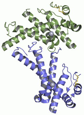
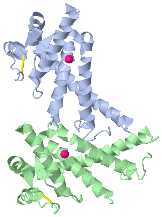
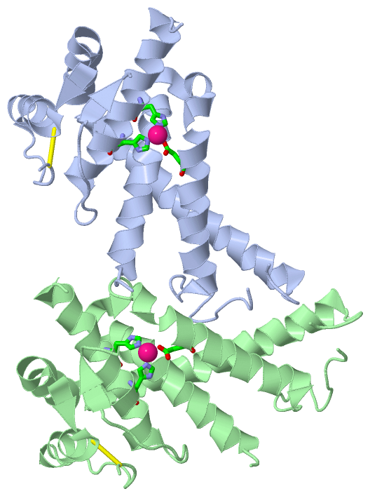
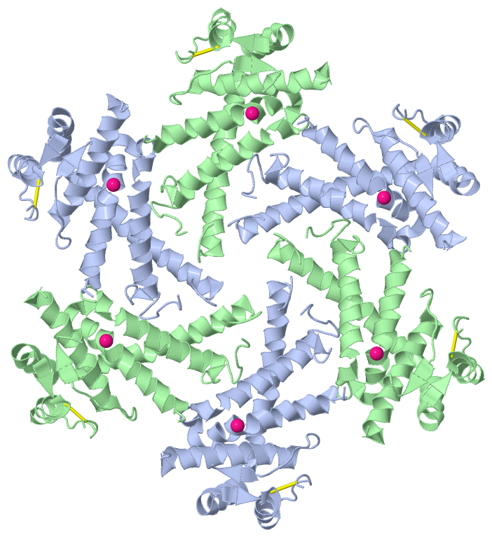
    GO:0046872    metal ion binding    Interacting selectively and non-covalently with any metal ion.
    GO:0004222    metalloendopeptidase activity    Catalysis of the hydrolysis of internal, alpha-peptide bonds in a polypeptide chain by a mechanism in which water acts as a nucleophile, one or two metal ions hold the water molecule in place, and charged amino acid side chains are ligands for the metal ions.
    GO:0008237    metallopeptidase activity    Catalysis of the hydrolysis of peptide bonds by a mechanism in which water acts as a nucleophile, one or two metal ions hold the water molecule in place, and charged amino acid side chains are ligands for the metal ions.
    GO:0000166    nucleotide binding    Interacting selectively and non-covalently with a nucleotide, any compound consisting of a nucleoside that is esterified with (ortho)phosphate or an oligophosphate at any hydroxyl group on the ribose or deoxyribose.
    GO:0008233    peptidase activity    Catalysis of the hydrolysis of a peptide bond. A peptide bond is a covalent bond formed when the carbon atom from the carboxyl group of one amino acid shares electrons with the nitrogen atom from the amino group of a second amino acid.
    GO:0008270    zinc ion binding    Interacting selectively and non-covalently with zinc (Zn) ions.
biological process
    GO:0030163    protein catabolic process    The chemical reactions and pathways resulting in the breakdown of a protein by the destruction of the native, active configuration, with or without the hydrolysis of peptide bonds.
    GO:0006508    proteolysis    The hydrolysis of proteins into smaller polypeptides and/or amino acids by cleavage of their peptide bonds.
cellular component
    GO:0016021    integral component of membrane    The component of a membrane consisting of the gene products and protein complexes having at least some part of their peptide sequence embedded in the hydrophobic region of the membrane.
    GO:0016020    membrane    A lipid bilayer along with all the proteins and protein complexes embedded in it an attached to it.
    GO:0005886    plasma membrane    The membrane surrounding a cell that separates the cell from its external environment. It consists of a phospholipid bilayer and associated proteins.

 Visualization

(-) Interactive Views

Asymmetric Unit
  Complete Structure
    Jena3D(integrated viewing of ligand, site, SAP, PROSITE, SCOP information)
    WebMol | AstexViewer[tm]@PDBe
(Java Applets, require no local installation except for Java; loading may be slow)
    STRAP
(Java WebStart application, automatic local installation, requires Java; full application with system access!)
    RasMol
(require local installation)
    Molscript (VRML)
(requires installation of a VRML viewer; select preferred view via VRML and generate a mono or stereo PDF format file)
 
  Ligands, Modified Residues, Ions
    HG  [ RasMol | Jena3D ]  +environment [ RasMol | Jena3D ]
 
  Sites
    AC1  [ RasMol ]  +environment [ RasMol ]
    AC2  [ RasMol ]  +environment [ RasMol ]
 
  Cis Peptide Bonds
(no "Cis Peptide Bonds" information available for 2di4)
 
Biological Unit
  Complete Structure
    Biological Unit 1  [ Jena3D ]

(-) Still Images

Jmol
  protein: cartoon or spacefill or dots and stick; nucleic acid: cartoon and stick; ligands: spacefill; active site: stick
Molscript
  protein, nucleic acid: cartoon; ligands: spacefill; active site: ball and stick

 Databases and Analysis Tools

(-) Databases

Access by PDB/NDB ID
  2di4
    Family and Domain InformationProDom | SYSTERS
    General Structural InformationGlycoscienceDB | MMDB | NDB | OCA | PDB | PDBe | PDBj | PDBsum | PDBWiki | PQS | PROTEOPEDIA
    Orientation in MembranesOPM
    Protein SurfaceSURFACE
    Secondary StructureDSSP (structure derived) | HSSP (homology derived)
    Structural GenomicsGeneCensus
    Structural NeighboursCE | VAST
    Structure ClassificationCATH | Dali | SCOP
    Validation and Original DataBMRB Data View | BMRB Restraints Grid | EDS | PROCHECK | RECOORD | WHAT_CHECK
 
Access by UniProt ID/Accession number
  FTSH_AQUAE | O67077
    Comparative Protein Structure ModelsModBase
    Genomic InformationEnsembl
    Protein-protein InteractionDIP
    Sequence, Family and Domain InformationInterPro | Pfam | SMART | UniProtKB/SwissProt
 
Access by Enzyme Classificator   (EC Number)
  3.4.24.-
    General Enzyme InformationBRENDA | EC-PDB | Enzyme | IntEnz
    PathwayKEGG | MetaCyc
 
Access by Disease Identifier   (MIM ID)
  (no 'MIM ID' available)
    Disease InformationOMIM
 
Access by GenAge ID
  (no 'GenAge ID' available)
    Age Related InformationGenAge

(-) Analysis Tools

Access by PDB/NDB ID
    Domain InformationXDom
    Interatomic Contacts of Structural UnitsCSU
    Ligand-protein ContactsLPC
    Protein CavitiescastP
    Sequence and Secondary StructurePDBCartoon
    Structure AlignmentSTRAP(Java WebStart application, automatic local installation, requires Java; full application with system access!)
    Structure and Sequence BrowserSTING
 
Access by UniProt ID/Accession number
  FTSH_AQUAE | O67077
    Protein Disorder PredictionDisEMBL | FoldIndex | GLOBPLOT (for more information see DisProt)

 Related Entries

(-) Entries Sharing at Least One Protein Chain (UniProt ID)

UniProtKB/Swiss-Prot
        FTSH_AQUAE | O670774ww0 4z8x

(-) Related Entries Specified in the PDB File

(no "Related Entries Specified in the PDB File" available for 2DI4)