| molecular function |
|---|
| | GO:0046789 | | host cell surface receptor binding | | Interacting selectively and non-covalently with a receptor on the host cell surface. |
| biological process |
|---|
| | GO:0075512 | | clathrin-dependent endocytosis of virus by host cell | | Any clathrin-mediated endocytosis that is involved in the uptake of a virus into a host cell. Begins by invagination of a specific region of the host cell plasma membrane around the bound virus to form a clathrin-coated pit, which then pinches off to form a clathrin-coated endocytic vesicle containing the virus. |
| | GO:0075509 | | endocytosis involved in viral entry into host cell | | Any endocytosis that is involved in the uptake of a virus into a host cell. |
| | GO:0039654 | | fusion of virus membrane with host endosome membrane | | Fusion of a virus membrane with a host endosome membrane. Occurs after internalization of the virus through the endosomal pathway, and results in release of the virus contents into the cell. |
| | GO:0019064 | | fusion of virus membrane with host plasma membrane | | Fusion of a viral membrane with the host cell membrane during viral entry. Results in release of the virion contents into the cytoplasm. |
| | GO:0039663 | | membrane fusion involved in viral entry into host cell | | Merging of the virion membrane and a host membrane (host plasma membrane or host organelle membrane) that is involved in the uptake of a virus into a host cell. |
| | GO:0019065 | | receptor-mediated endocytosis of virus by host cell | | Any receptor-mediated endocytosis that is involved in the uptake of a virus into a host cell; successive instances of virus endocytosis result in the accumulation of virus particles within the cell. |
| | GO:0046718 | | viral entry into host cell | | The process that occurs after viral attachment by which a virus, or viral nucleic acid, breaches the plasma membrane or cell envelope and enters the host cell. The process ends when the viral nucleic acid is released into the host cell cytoplasm. |
| | GO:0016032 | | viral process | | A multi-organism process in which a virus is a participant. The other participant is the host. Includes infection of a host cell, replication of the viral genome, and assembly of progeny virus particles. In some cases the viral genetic material may integrate into the host genome and only subsequently, under particular circumstances, 'complete' its life cycle. |
| | GO:0019062 | | virion attachment to host cell | | The process by which a virion protein binds to molecules on the host cellular surface or host cell surface projection. |
| cellular component |
|---|
| | GO:0033644 | | host cell membrane | | Double layer of lipid molecules as it encloses host cells, and, in eukaryotes, many organelles; may be a single or double lipid bilayer; also includes associated proteins. The host is defined as the larger of the organisms involved in a symbiotic interaction. |
| | GO:0020002 | | host cell plasma membrane | | The plasma membrane surrounding a host cell. |
| | GO:0016021 | | integral component of membrane | | The component of a membrane consisting of the gene products and protein complexes having at least some part of their peptide sequence embedded in the hydrophobic region of the membrane. |
| | GO:0016020 | | membrane | | A lipid bilayer along with all the proteins and protein complexes embedded in it an attached to it. |
| | GO:0019031 | | viral envelope | | The lipid bilayer of a virion that surrounds the protein capsid. May also contain glycoproteins. |
| | GO:0019012 | | virion | | The complete fully infectious extracellular virus particle. |
| | GO:0055036 | | virion membrane | | The lipid bilayer surrounding a virion. |
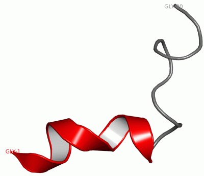
 Description
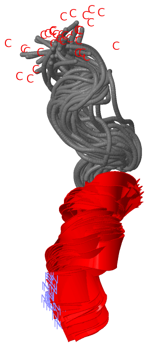