Show PDB file:   
         Plain Text   HTML   (compressed file size)
QuickSearch:   
by PDB,NDB,UniProt,PROSITE Code or Search Term(s)  
(-)NMR Structure - manually
(-)NMR Structure - model 1
(-)NMR Structure - model 1, sites
(-)NMR Structure - all models
collapse expand < >
Image NMR Structure - manually
NMR Structure - manually  (Jmol Viewer)
Image NMR Structure - model 1
NMR Structure - model 1  (Jmol Viewer)
Image NMR Structure - model 1, sites
NMR Structure - model 1, sites  (Jmol Viewer)
Image NMR Structure - all models
NMR Structure - all models  (Jmol Viewer)

(-) Description

Title :  THE SOLUTION STRUCTURE OF THE MYND DOMAIN (LEU384-CYS430) OF HUMAN ZINC FINGER MYND DOMAIN CONTAINING PROTEIN 10
 
Authors :  A. Sasagawa, K. Miyamoto, N. Tochio, S. Koshiba, M. Inoue, T. Kigawa, S. Yokoyama, Riken Structural Genomics/Proteomics Initiative (Rsgi)
Date :  14 Dec 05  (Deposition) - 14 Jun 06  (Release) - 24 Feb 09  (Revision)
Method :  SOLUTION NMR
Resolution :  NOT APPLICABLE
Chains :  NMR Structure  :  A  (20x)
Keywords :  Zf-Mynd, Blu Protein, C3H, Structural Genomics, Nppsfa, National Project On Protein Structural And Functional Analyses, Riken Structural Genomics/Proteomics Initiative, Rsgi, Metal Binding Protein (Keyword Search: [Gene Ontology, PubMed, Web (Google))
 
Reference :  A. Sasagawa, K. Miyamoto, N. Tochio, S. Koshiba, M. Inoue, T. Kigawa, S. Yokoyama
The Solution Structure Of The Mynd Domain (Leu384-Cys430) Of Human Zinc Finger Mynd Domain Containing Protein 10
To Be Published
PubMed: search
(for further references see the PDB file header)

(-) Compounds

Molecule 1 - ZINC FINGER MYND DOMAIN CONTAINING PROTEIN 10
    ChainsA
    EngineeredYES
    Expression SystemCELL FREE SYNTHESIS
    Expression System PlasmidP050302-43
    Expression System Vector TypePLASMID
    FragmentMYND DOMAIN
    GeneZMYND10
    Organism CommonHUMAN
    Organism ScientificHOMO SAPIENS
    Organism Taxid9606
    Other DetailsCELL-FREE PROTEIN SYNTHESIS
    SynonymBLU PROTEIN

 Structural Features

(-) Chains, Units

  
NMR Structure (20x)

Summary Information (see also Sequences/Alignments below)

(-) Ligands, Modified Residues, Ions  (1, 2)

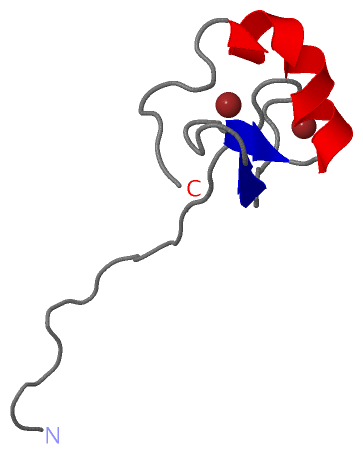
NMR Structure (1, 2)
No.NameCountTypeFull Name
1ZN2Ligand/IonZINC ION

(-) Sites  (2, 2)

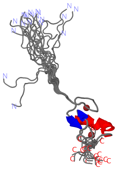
NMR Structure (2, 2)
No.NameEvidenceResiduesDescription
1AC1SOFTWARECYS A:29 , ARG A:31 , CYS A:32 , HIS A:50 , CYS A:54BINDING SITE FOR RESIDUE ZN A 201
2AC2SOFTWARECYS A:18 , CYS A:21 , CYS A:38 , CYS A:42BINDING SITE FOR RESIDUE ZN A 401

(-) SS Bonds  (0, 0)

(no "SS Bond" information available for 2DAN)

(-) Cis Peptide Bonds  (0, 0)

(no "Cis Peptide Bond" information available for 2DAN)

 Sequence-Structure Mapping

(-) SAPs(SNPs)/Variants  (1, 1)

NMR Structure (1, 1)
  dbSNPPDB
No.SourceVariant IDVariantUniProt IDStatusIDChainVariant
1UniProtVAR_014227R407QZMY10_HUMANUnclassified182064110AR31Q

  SNP/SAP Summary Statistics (UniProtKB/Swiss-Prot)

(-) PROSITE Motifs  (2, 1)

NMR Structure (2, 1)
 PROSITEUniProtKBPDB
No.IDACDescriptionIDLocationCountLocation
1ZF_MYND_1PS01360 Zinc finger MYND-type signature.ZMY10_HUMAN394-430  1A:18-54
2ZF_MYND_2PS50865 Zinc finger MYND-type profile.ZMY10_HUMAN394-430  1A:18-54

(-) Exons   (3, 3)

NMR Structure (3, 3)
 ENSEMBLUniProtKBPDB
No.Transcript IDExonExon IDGenome LocationLengthIDLocationLengthCountLocationLength
1.1aENST000002317491aENSE00001372499chr3:50384283-503829191365ZMY10_HUMAN1-31310--
1.2bENST000002317492bENSE00001785120chr3:50382663-50382555109ZMY10_HUMAN31-67370--
1.4bENST000002317494bENSE00001684837chr3:50381281-50381165117ZMY10_HUMAN68-106390--
1.5ENST000002317495ENSE00001803532chr3:50381047-5038099454ZMY10_HUMAN107-124180--
1.6bENST000002317496bENSE00001806257chr3:50380875-50380738138ZMY10_HUMAN125-170460--
1.6dENST000002317496dENSE00001731202chr3:50380637-5038054989ZMY10_HUMAN171-200300--
1.6fENST000002317496fENSE00001797495chr3:50380449-50380349101ZMY10_HUMAN200-234350--
1.6iENST000002317496iENSE00001598571chr3:50380000-50379828173ZMY10_HUMAN234-291580--
1.6lENST000002317496lENSE00001654103chr3:50379571-50379446126ZMY10_HUMAN292-333420--
1.7ENST000002317497ENSE00000860731chr3:50379362-50379241122ZMY10_HUMAN334-374411A:1-7 (gaps)21
1.8ENST000002317498ENSE00001610366chr3:50379130-50379005126ZMY10_HUMAN374-416431A:8-4033
1.9cENST000002317499cENSE00001917579chr3:50378916-50378541376ZMY10_HUMAN416-440251A:40-6021

(-) Sequences/Alignments

NMR Structure
   Reformat: Number of residues per line =  ('0' or empty: single-line sequence representation)
  Number of residues per labelling interval =   
  UniProt sequence: complete  aligned part    
   Show mapping: SCOP domains CATH domains Pfam domains Secondary structure (by author)
SAPs(SNPs) PROSITE motifs Exons
(details for a mapped element are shown in a popup box when the mouse pointer rests over it)
Chain A from PDB  Type:PROTEIN  Length:60
 aligned with ZMY10_HUMAN | O75800 from UniProtKB/Swiss-Prot  Length:440

    Alignment length:93
                                   353       363       373       383       393       403       413       423       433   
          ZMY10_HUMAN   344 ENRGKWQAIAKHQLQHVFSPSEQDLRLQARRWAETYRLDVLEAVAPERPRCAYCSAEASKRCSRCQNEWYCCRECQVKHWEKHGKTCVLAAQG 436
               SCOP domains ----------------------------------------d2dana1 A:8-54                                 ------ SCOP domains
               CATH domains --------------------------------------------------------------------------------------------- CATH domains
               Pfam domains --------------------------------------------------------------------------------------------- Pfam domains
         Sec.struct. author ....--------------...-------------------...................ee.......ee.hhhhhhhhhhhhh......... Sec.struct. author
                 SAPs(SNPs) ---------------------------------------------------------------Q----------------------------- SAPs(SNPs)
                PROSITE (1) --------------------------------------------------ZF_MYND_1  PDB: A:18-54              ------ PROSITE (1)
                PROSITE (2) --------------------------------------------------ZF_MYND_2  PDB: A:18-54              ------ PROSITE (2)
           Transcript 1 (1) Exon 1.7  PDB: A:1-7 (gaps)    -----------------------------------------Exon 1.9c             Transcript 1 (1)
           Transcript 1 (2) ------------------------------Exon 1.8  PDB: A:8-40 UniProt: 374-416     -------------------- Transcript 1 (2)
                 2dan A   1 GSSG--------------SSG-------------------LEAVAPERPRCAYCSAEASKRCSRCQNEWYCCRECQVKHWEKHGKTCSGPSSG  60
                               |     -        |6|        -         -|       17        27        37        47        57   
                               4              5 7                   8                                                    

   Legend:   → Mismatch (orange background)
  - → Gap (green background, '-', border residues have a numbering label)
    → Modified Residue (blue background, lower-case, 'x' indicates undefined single-letter code, labelled with number + name)
  x → Chemical Group (purple background, 'x', labelled with number + name, e.g. ACE or NH2)
  extra numbering lines below/above indicate numbering irregularities and modified residue names etc., number ends below/above '|'

 Classification and Annotation

(-) SCOP Domains  (1, 1)

NMR Structure

(-) CATH Domains  (0, 0)

(no "CATH Domain" information available for 2DAN)

(-) Pfam Domains  (0, 0)

(no "Pfam Domain" information available for 2DAN)

(-) Gene Ontology  (9, 9)

NMR Structure(hide GO term definitions)
Chain A   (ZMY10_HUMAN | O75800)
molecular function
    GO:0046872    metal ion binding    Interacting selectively and non-covalently with any metal ion.
    GO:0005515    protein binding    Interacting selectively and non-covalently with any protein or protein complex (a complex of two or more proteins that may include other nonprotein molecules).
biological process
    GO:0036159    inner dynein arm assembly    The aggregation, arrangement and bonding together of a set of components to form an axonemal dynein inner arm, an inner arm structure present on the outer doublet microtubules of ciliary and flagellar axonemes.
    GO:0044458    motile cilium assembly    The aggregation, arrangement and bonding together of a set of components to form a motile cilium.
    GO:0036158    outer dynein arm assembly    The aggregation, arrangement and bonding together of a set of components to form an axonemal dynein outer arm, an outer arm structure present on the outer doublet microtubules of ciliary and flagellar axonemes.
cellular component
    GO:0034451    centriolar satellite    A small (70-100 nm) cytoplasmic granule that contains a number of centrosomal proteins; centriolar satellites traffic toward microtubule minus ends and are enriched near the centrosome.
    GO:0005737    cytoplasm    All of the contents of a cell excluding the plasma membrane and nucleus, but including other subcellular structures.
    GO:0005856    cytoskeleton    Any of the various filamentous elements that form the internal framework of cells, and typically remain after treatment of the cells with mild detergent to remove membrane constituents and soluble components of the cytoplasm. The term embraces intermediate filaments, microfilaments, microtubules, the microtrabecular lattice, and other structures characterized by a polymeric filamentous nature and long-range order within the cell. The various elements of the cytoskeleton not only serve in the maintenance of cellular shape but also have roles in other cellular functions, including cellular movement, cell division, endocytosis, and movement of organelles.
    GO:0005815    microtubule organizing center    An intracellular structure that can catalyze gamma-tubulin-dependent microtubule nucleation and that can anchor microtubules by interacting with their minus ends, plus ends or sides.

 Visualization

(-) Interactive Views

NMR Structure
  Complete Structure
    Jena3D(integrated viewing of ligand, site, SAP, PROSITE, SCOP information)
    WebMol | AstexViewer[tm]@PDBe
(Java Applets, require no local installation except for Java; loading may be slow)
    STRAP
(Java WebStart application, automatic local installation, requires Java; full application with system access!)
    RasMol
(require local installation)
    Molscript (VRML)
(requires installation of a VRML viewer; select preferred view via VRML and generate a mono or stereo PDF format file)
 
  Ligands, Modified Residues, Ions
    ZN  [ RasMol | Jena3D ]  +environment [ RasMol | Jena3D ]
 
  Sites
    AC1  [ RasMol ]  +environment [ RasMol ]
    AC2  [ RasMol ]  +environment [ RasMol ]
 
  Cis Peptide Bonds
(no "Cis Peptide Bonds" information available for 2dan)
 

(-) Still Images

Jmol
  protein: cartoon or spacefill or dots and stick; nucleic acid: cartoon and stick; ligands: spacefill; active site: stick
Molscript
  protein, nucleic acid: cartoon; ligands: spacefill; active site: ball and stick

 Databases and Analysis Tools

(-) Databases

Access by PDB/NDB ID
  2dan
    Family and Domain InformationProDom | SYSTERS
    General Structural InformationGlycoscienceDB | MMDB | NDB | OCA | PDB | PDBe | PDBj | PDBsum | PDBWiki | PQS | PROTEOPEDIA
    Orientation in MembranesOPM
    Protein SurfaceSURFACE
    Secondary StructureDSSP (structure derived) | HSSP (homology derived)
    Structural GenomicsGeneCensus
    Structural NeighboursCE | VAST
    Structure ClassificationCATH | Dali | SCOP
    Validation and Original DataBMRB Data View | BMRB Restraints Grid | EDS | PROCHECK | RECOORD | WHAT_CHECK
 
Access by UniProt ID/Accession number
  ZMY10_HUMAN | O75800
    Comparative Protein Structure ModelsModBase
    Genomic InformationEnsembl
    Protein-protein InteractionDIP
    Sequence, Family and Domain InformationInterPro | Pfam | SMART | UniProtKB/SwissProt
 
Access by Enzyme Classificator   (EC Number)
  (no 'Enzyme Classificator' available)
    General Enzyme InformationBRENDA | EC-PDB | Enzyme | IntEnz
    PathwayKEGG | MetaCyc
 
Access by Disease Identifier   (MIM ID)
  (no 'MIM ID' available)
    Disease InformationOMIM
 
Access by GenAge ID
  (no 'GenAge ID' available)
    Age Related InformationGenAge

(-) Analysis Tools

Access by PDB/NDB ID
    Domain InformationXDom
    Interatomic Contacts of Structural UnitsCSU
    Ligand-protein ContactsLPC
    Protein CavitiescastP
    Sequence and Secondary StructurePDBCartoon
    Structure AlignmentSTRAP(Java WebStart application, automatic local installation, requires Java; full application with system access!)
    Structure and Sequence BrowserSTING
 
Access by UniProt ID/Accession number
  ZMY10_HUMAN | O75800
    Protein Disorder PredictionDisEMBL | FoldIndex | GLOBPLOT (for more information see DisProt)

 Related Entries

(-) Entries Sharing at Least One Protein Chain (UniProt ID)

UniProtKB/Swiss-Prot
        ZMY10_HUMAN | O758002d8q

(-) Related Entries Specified in the PDB File

2d8q THE SAME PROTEIN DOMAIN OF THE DIFFERENT LENGTH RELATED ID: HSS001004006.1 RELATED DB: TARGETDB