| molecular function |
|---|
| | GO:0031681 | | G-protein beta-subunit binding | | Interacting selectively and non-covalently with a G-protein beta subunit. |
| | GO:0005096 | | GTPase activator activity | | Binds to and increases the activity of a GTPase, an enzyme that catalyzes the hydrolysis of GTP. |
| | GO:0004871 | | signal transducer activity | | Conveys a signal across a cell to trigger a change in cell function or state. A signal is a physical entity or change in state that is used to transfer information in order to trigger a response. |
| biological process |
|---|
| | GO:0007186 | | G-protein coupled receptor signaling pathway | | A series of molecular signals that proceeds with an activated receptor promoting the exchange of GDP for GTP on the alpha-subunit of an associated heterotrimeric G-protein complex. The GTP-bound activated alpha-G-protein then dissociates from the beta- and gamma-subunits to further transmit the signal within the cell. The pathway begins with receptor-ligand interaction, or for basal GPCR signaling the pathway begins with the receptor activating its G protein in the absence of an agonist, and ends with regulation of a downstream cellular process, e.g. transcription. The pathway can start from the plasma membrane, Golgi or nuclear membrane (PMID:24568158 and PMID:16902576). |
| | GO:0035556 | | intracellular signal transduction | | The process in which a signal is passed on to downstream components within the cell, which become activated themselves to further propagate the signal and finally trigger a change in the function or state of the cell. |
| | GO:0009968 | | negative regulation of signal transduction | | Any process that stops, prevents, or reduces the frequency, rate or extent of signal transduction. |
| | GO:0043547 | | positive regulation of GTPase activity | | Any process that activates or increases the activity of a GTPase. |
| | GO:0006457 | | protein folding | | The process of assisting in the covalent and noncovalent assembly of single chain polypeptides or multisubunit complexes into the correct tertiary structure. |
| cellular component |
|---|
| | GO:0005737 | | cytoplasm | | All of the contents of a cell excluding the plasma membrane and nucleus, but including other subcellular structures. |
| | GO:0005829 | | cytosol | | The part of the cytoplasm that does not contain organelles but which does contain other particulate matter, such as protein complexes. |
| | GO:0005834 | | heterotrimeric G-protein complex | | Any of a family of heterotrimeric GTP-binding and hydrolyzing proteins; they belong to a superfamily of GTPases that includes monomeric proteins such as EF-Tu and RAS. Heterotrimeric G-proteins consist of three subunits; the alpha subunit contains the guanine nucleotide binding site and possesses GTPase activity; the beta and gamma subunits are tightly associated and function as a beta-gamma heterodimer; extrinsic plasma membrane proteins (cytoplasmic face) that function as a complex to transduce signals from G-protein coupled receptors to an effector protein. |
| | GO:0016020 | | membrane | | A lipid bilayer along with all the proteins and protein complexes embedded in it an attached to it. |
| | GO:0005886 | | plasma membrane | | The membrane surrounding a cell that separates the cell from its external environment. It consists of a phospholipid bilayer and associated proteins. |
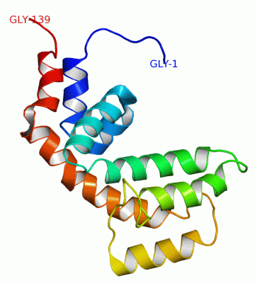
 Description
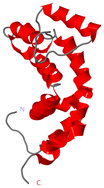
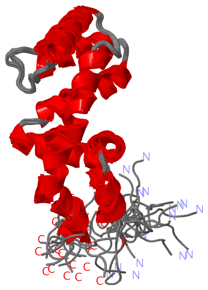