|
|
|
|
 Description
|
|
Compounds
|
||||||||||||||||||||
Chains, Units
Summary Information (see also Sequences/Alignments below) |
Ligands, Modified Residues, Ions (2, 12)| Asymmetric Unit (2, 12) Biological Unit 1 (1, 1) Biological Unit 2 (1, 1) |
Sites (11, 11)
Asymmetric Unit (11, 11)
|
SS Bonds (2, 2)
Asymmetric Unit
|
||||||||||||
Cis Peptide Bonds (0, 0)| (no "Cis Peptide Bond" information available for 2D0W) |
SAPs(SNPs)/Variants (0, 0)| (no "SAP(SNP)/Variant" information available for 2D0W) |
PROSITE Motifs (0, 0)| (no "PROSITE Motif" information available for 2D0W) |
Exons (0, 0)| (no "Exon" information available for 2D0W) |
Sequences/Alignments
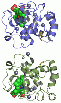
Asymmetric UnitChain A from PDB Type:PROTEIN Length:168 aligned with Q4AE24_9RHIZ | Q4AE24 from UniProtKB/TrEMBL Length:193 Alignment length:168 33 43 53 63 73 83 93 103 113 123 133 143 153 163 173 183 Q4AE24_9RHIZ 24 AQEVFRNTVTGEALDVEGQAPKEGRDTPAVKQFMQTGVDPYVEVAGCLPKGEEIYLESCSGCHGHIGEGKVGPGLNDSYWTYPKNTTDKGLFETIFGGANGMMGPHGQDLELDNMLKLIAWIRHIQKDDVADADWLSDEQKKNFKPFDIKAWEATGKAAAEKAQCKIS 191 SCOP domains d2d0wa_ A: automated matches SCOP domains CATH domains 2d0wA00 A:1-168 Cytochrome c CATH domains Pfam domains ------------------------------------------------------------------------------------------------------------------------------------------------------------------------ Pfam domains SAPs(SNPs) ------------------------------------------------------------------------------------------------------------------------------------------------------------------------ SAPs(SNPs) PROSITE ------------------------------------------------------------------------------------------------------------------------------------------------------------------------ PROSITE Transcript ------------------------------------------------------------------------------------------------------------------------------------------------------------------------ Transcript 2d0w A 1 AQEVFRNTVTGEALDVEGQAPKEGRDTPAVKQFMQTGVDPYVEVAGCLPKGEEIYLESCSGCHGHIGEGKVGPGLNDSYWTYPKNTTDKGLFETIFGGANGMMGPHGQDLELDNMLKLIAWIRHIQKDDVADADWLSDEQKKNFKPFDIKAWEATGKAAAEKAQCKIS 168 10 20 30 40 50 60 70 80 90 100 110 120 130 140 150 160 Chain B from PDB Type:PROTEIN Length:168 aligned with Q4AE24_9RHIZ | Q4AE24 from UniProtKB/TrEMBL Length:193 Alignment length:168 33 43 53 63 73 83 93 103 113 123 133 143 153 163 173 183 Q4AE24_9RHIZ 24 AQEVFRNTVTGEALDVEGQAPKEGRDTPAVKQFMQTGVDPYVEVAGCLPKGEEIYLESCSGCHGHIGEGKVGPGLNDSYWTYPKNTTDKGLFETIFGGANGMMGPHGQDLELDNMLKLIAWIRHIQKDDVADADWLSDEQKKNFKPFDIKAWEATGKAAAEKAQCKIS 191 SCOP domains d2d0wb_ B: automated matches SCOP domains CATH domains 2d0wB00 B:1-168 Cytochrome c CATH domains Pfam domains ------------------------------------------------------------------------------------------------------------------------------------------------------------------------ Pfam domains SAPs(SNPs) ------------------------------------------------------------------------------------------------------------------------------------------------------------------------ SAPs(SNPs) PROSITE ------------------------------------------------------------------------------------------------------------------------------------------------------------------------ PROSITE Transcript ------------------------------------------------------------------------------------------------------------------------------------------------------------------------ Transcript 2d0w B 1 AQEVFRNTVTGEALDVEGQAPKEGRDTPAVKQFMQTGVDPYVEVAGCLPKGEEIYLESCSGCHGHIGEGKVGPGLNDSYWTYPKNTTDKGLFETIFGGANGMMGPHGQDLELDNMLKLIAWIRHIQKDDVADADWLSDEQKKNFKPFDIKAWEATGKAAAEKAQCKIS 168 10 20 30 40 50 60 70 80 90 100 110 120 130 140 150 160
|
||||||||||||||||||||
SCOP Domains (1, 2)
Asymmetric Unit
 |
CATH Domains (1, 2)
Asymmetric Unit
|
Pfam Domains (0, 0)| (no "Pfam Domain" information available for 2D0W) |
Gene Ontology (7, 7)|
Asymmetric Unit(hide GO term definitions) Chain A,B (Q4AE24_9RHIZ | Q4AE24)
|
||||||||||||||||||||||||||||||||||||||||||||||||||||||||||||
Interactive Views
|
||||||||||||||||||||||||||||||||||||||||||||||||||||||||||||||||||||||||||||||||||||||||||||||||||||||||||||||||||||||||||||||||||||||||||||||||||||||||||||||||||||||||||||||||||||||||||||||||||||||||||||||||||||||||||
Still Images
|
||||||||||||||||
Databases
|
||||||||||||||||||||||||||||||||||||||||||||||||||||||||||||||||||||||||||||||||||||||||||||||||||||||||||||||||||||||||||||||||||||||||||||||||||||||||||||||||
Analysis Tools
|
|||||||||||||||||||||||||||||||||||||||||||||||||||||||||||||
Entries Sharing at Least One Protein Chain (UniProt ID)
Related Entries Specified in the PDB File
|
|