|
|
|
|
 Description
|
|
Compounds
|
||||||||||||||||||||||||||||||||||||||||||||||||
Chains, Units
Summary Information (see also Sequences/Alignments below) |
Ligands, Modified Residues, Ions (0, 0)| (no "Ligand,Modified Residues,Ions" information available for 2CZN) |
Sites (0, 0)| (no "Site" information available for 2CZN) |
SS Bonds (0, 0)| (no "SS Bond" information available for 2CZN) |
Cis Peptide Bonds (0, 0)| (no "Cis Peptide Bond" information available for 2CZN) |
SAPs(SNPs)/Variants (0, 0)| (no "SAP(SNP)/Variant" information available for 2CZN) |
PROSITE Motifs (0, 0)| (no "PROSITE Motif" information available for 2CZN) |
Exons (0, 0)| (no "Exon" information available for 2CZN) |
Sequences/Alignments
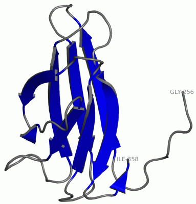
NMR StructureChain A from PDB Type:PROTEIN Length:103 aligned with Q8U1H5_PYRFU | Q8U1H5 from UniProtKB/TrEMBL Length:717 Alignment length:103 265 275 285 295 305 315 325 335 345 355 Q8U1H5_PYRFU 256 TTTTPVPVSGSLEVKVNDWGSGAEYDVTLNLDGQYDWTVKVKLAPGATVGSFWSANKQEGNGYVIFTPVSWNKGPTATFGFIVNGPQGDKVEEITLEINGQVI 358 SCOP domains ------------------------------------------------------------------------------------------------------- SCOP domains CATH domains ------------------------------------------------------------------------------------------------------- CATH domains Pfam domains ------------------------------------------------------------------------------------------------------- Pfam domains SAPs(SNPs) ------------------------------------------------------------------------------------------------------- SAPs(SNPs) PROSITE ------------------------------------------------------------------------------------------------------- PROSITE Transcript ------------------------------------------------------------------------------------------------------- Transcript 2czn A 256 GPTTPVPVSGSLEVKVNDWGSGAEYDVTLNLDGQYDWTVKVKLAPGATVGSFWSANKQEGNGYVIFTPVSWNKGPTATFGFIVNGPQGDKVEEITLEINGQVI 358 265 275 285 295 305 315 325 335 345 355
|
||||||||||||||||||||
SCOP Domains (0, 0)| (no "SCOP Domain" information available for 2CZN) |
CATH Domains (0, 0)| (no "CATH Domain" information available for 2CZN) |
Pfam Domains (0, 0)| (no "Pfam Domain" information available for 2CZN) |
Gene Ontology (6, 6)|
NMR Structure(hide GO term definitions) Chain A (Q8U1H5_PYRFU | Q8U1H5)
|
||||||||||||||||||||||||||||||||||||||||||||||||
Interactive Views
|
||||||||||||||||||||||||||||||||||||||||||||||||||||||||||||||||||||||||||||||||||||||||||||||||||||||||||||||||||||
Still Images
|
||||||||||||||||
Databases
|
||||||||||||||||||||||||||||||||||||||||||||||||||||||||||||||||||||||||||||||||||||||||||||||||||||||||||||||||||||||||||||||||||||||||||||||||||||||||||||||||
Analysis Tools
|
|||||||||||||||||||||||||||||||||||||||||||||||||||||||||||||
Entries Sharing at Least One Protein Chain (UniProt ID)
Related Entries Specified in the PDB File
|
|