Show PDB file:   
         Plain Text   HTML   (compressed file size)
QuickSearch:   
by PDB,NDB,UniProt,PROSITE Code or Search Term(s)  
(-)NMR Structure - manually
(-)NMR Structure - model 1
(-)NMR Structure - model 1, sites
(-)NMR Structure - all models
collapse expand < >
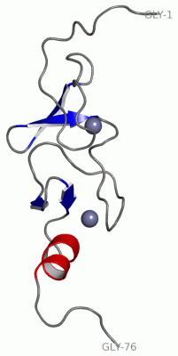
Image NMR Structure - manually
NMR Structure - manually  (Jmol Viewer)
Image NMR Structure - model 1
NMR Structure - model 1  (Jmol Viewer)
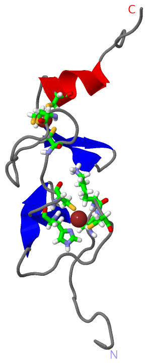
Image NMR Structure - model 1, sites
NMR Structure - model 1, sites  (Jmol Viewer)
Image NMR Structure - all models
NMR Structure - all models  (Jmol Viewer)

(-) Description

Title :  SOLUTION STRUCTURE OF THE LIM DOMAIN OF HUMAN CYSTEINE-RICH PROTEIN 2
 
Authors :  M. Yoneyama, A. Sasagawa, T. Tomizawa, N. Tochio, S. Koshiba, M. Inoue, T. Kigawa, S. Yokoyama, Riken Structural Genomics/Proteomics Initiative (Rsgi)
Date :  25 May 05  (Deposition) - 25 Nov 05  (Release) - 24 Feb 09  (Revision)
Method :  SOLUTION NMR
Resolution :  NOT APPLICABLE
Chains :  NMR Structure  :  A  (20x)
Keywords :  Crp2, Crip2, Esp1 Protein, Zinc-Binding, Structural Genomics, Nppsfa, National Project On Protein Structural And Functional Analyses, Riken Structural Genomics/Proteomics Initiative, Rsgi, Metal Binding Protein (Keyword Search: [Gene Ontology, PubMed, Web (Google)] )
 
Reference :  M. Yoneyama, A. Sasagawa, T. Tomizawa, N. Tochio, S. Koshiba, M. Inoue, T. Kigawa, S. Yokoyama
Solution Structure Of The Lim Domain Of Human Cysteine-Rich Protein 2
To Be Published
PubMed: search
(for further references see the PDB file header)

(-) Compounds

Molecule 1 - CYSTEINE-RICH PROTEIN 2
    Chains: A
    Engineered: YES
    Expression System Plasmid: P040614-03
    Expression System Vector Type: PLASMID
    Fragment: LIM DOMAIN
    Gene: CRIP2
    Organism Common: HUMAN
    Organism Scientific: HOMO SAPIENS
    Organism Taxid: 9606
    Other Details: CELL-FREE PROTEIN SYNTHESIS
    Synonym: CRP2, ESP1 PROTEIN

 Structural Features

(-) Chains, Units

  
NMR Structure (20x): 

Summary Information (see also Sequences/Alignments below)

(-) Ligands, Modified Residues, Ions  (1, 2)

NMR Structure (1, 2)
No.NameCountTypeFull Name
1ZN2Ligand/IonZINC ION

(-) Sites  (2, 2)

NMR Structure (2, 2)
No.NameEvidenceResiduesDescription
1AC1SOFTWARECYS A:12 , LYS A:14 , CYS A:15 , HIS A:33 , CYS A:36BINDING SITE FOR RESIDUE ZN A 201
2AC2SOFTWARECYS A:39 , CYS A:42 , CYS A:60 , CYS A:64BINDING SITE FOR RESIDUE ZN A 401

(-) SS Bonds  (0, 0)

(no "SS Bond" information available for 2CU8)

(-) Cis Peptide Bonds  (1, 20)

NMR Structure
No.ModelResidues
11, 2, 3, 4, 5, 6, 7, 8, 9, 10, 11, 12, 13, 14, 15, 16, 17, 18, 19, 20Lys A:62 -Pro A:63

 Sequence-Structure Mapping

(-) SAPs(SNPs)/Variants  (0, 0)

(no "SAP(SNP)/Variant" information available for 2CU8)

(-) PROSITE Motifs  (2, 2)

NMR Structure (2, 2)
 PROSITEUniProtKBPDB
No.IDACDescriptionIDLocationCountLocation
1LIM_DOMAIN_2PS50023 LIM domain profile.CRIP2_HUMAN3-64
124-185
  1A:10-70
-
2LIM_DOMAIN_1PS00478 LIM zinc-binding domain signature.CRIP2_HUMAN5-39
126-160
  1A:12-46
-

(-) Exons   (4, 4)

NMR Structure (4, 4)
 ENSEMBLUniProtKBPDB
No.Transcript IDExonExon IDGenome LocationLengthIDLocationLengthCountLocationLength
1.2ENST000003291462ENSE00001305445chr14:105940960-105941242283CRIP2_HUMAN1-15151A:8-2215
1.3ENST000003291463ENSE00001293412chr14:105944603-10594469795CRIP2_HUMAN15-46321A:22-5332
1.4aENST000003291464aENSE00001669917chr14:105944787-10594484458CRIP2_HUMAN47-66201A:54-7017
1.5ENST000003291465ENSE00001320233chr14:105945068-105945208141CRIP2_HUMAN66-113481A:71-766
1.6ENST000003291466ENSE00001307611chr14:105945316-10594538469CRIP2_HUMAN113-136240--
1.7ENST000003291467ENSE00001326756chr14:105945462-10594555695CRIP2_HUMAN136-167320--
1.8ENST000003291468ENSE00001687544chr14:105945773-10594583058CRIP2_HUMAN168-187200--
1.9bENST000003291469bENSE00001312310chr14:105945923-105946507585CRIP2_HUMAN187-208220--

(-) Sequences/Alignments

NMR Structure
   Reformat: Number of residues per line =  ('0' or empty: single-line sequence representation)
  Number of residues per labelling interval =   
  UniProt sequence: complete  aligned part    
   Show mapping: SCOP domains CATH domains Pfam domains Secondary structure (by author)
SAPs(SNPs) PROSITE motifs Exons
(details for a mapped element are shown in a popup box when the mouse pointer rests over it)
Chain A from PDB  Type:PROTEIN  Length:76
 aligned with CRIP2_HUMAN | P52943 from UniProtKB/Swiss-Prot  Length:208

    Alignment length:116
                                   1                                                                                                            
                                   | 3        13        23        33        43        53        63        73        83        93       103      
          CRIP2_HUMAN     - -------MASKCPKCDKTVYFAEKVSSLGKDWHKFCLKCERCSKTLTPGGHAEHDGKPFCHKPCYATLFGPKGVNIGGAGSYIYEKPLAEGPQVTGPIEVPAARAEERKASGPPKG 109
               SCOP domains -------d2cu8a1 A:8-37                d2cu8a2 A:38-70                  ---------------------------------------------- SCOP domains
               CATH domains 2cu8A00 A:1-76 Cysteine Rich Protein                                                                                 CATH domains
               Pfam domains -------------------------------------------------------------------------------------------------------------------- Pfam domains
         Sec.struct. author .......................eeee..eeee..................eee..eee...hhhhhhh.----------------------------------------...... Sec.struct. author
                 SAPs(SNPs) -------------------------------------------------------------------------------------------------------------------- SAPs(SNPs)
                PROSITE (1) ---------LIM_DOMAIN_2  PDB: A:10-70 UniProt: 3-64                      --------------------------------------------- PROSITE (1)
                PROSITE (2) -----------LIM_DOMAIN_1  PDB: A:12-46         ---------------------------------------------------------------------- PROSITE (2)
           Transcript 1 (1) -------Exon 1.2       -------------------------------Exon 1.4a           ------------------------------------------- Transcript 1 (1)
           Transcript 1 (2) ---------------------Exon 1.3  PDB: A:22-53          -------------------Exon 1.5  PDB: A:71-76 UniProt: 66-113       Transcript 1 (2)
                 2cu8 A   1 GSSGSSGMASKCPKCDKTVYFAEKVSSLGKDWHKFCLKCERCSKTLTPGGHAEHDGKPFCHKPCYATLFG----------------------------------------SGPSSG  76
                                    10        20        30        40        50        60        70         -         -         -         -|     
                                                                                                70                                       71     

   Legend:   → Mismatch (orange background)
  - → Gap (green background, '-', border residues have a numbering label)
    → Modified Residue (blue background, lower-case, 'x' indicates undefined single-letter code, labelled with number + name)
  x → Chemical Group (purple background, 'x', labelled with number + name, e.g. ACE or NH2)
  extra numbering lines below/above indicate numbering irregularities and modified residue names etc., number ends below/above '|'

 Classification and Annotation

(-) SCOP Domains  (1, 2)

NMR Structure

(-) CATH Domains  (1, 1)

NMR Structure
(-)
Class: Mainly Beta (13760)

(-) Pfam Domains  (0, 0)

(no "Pfam Domain" information available for 2CU8)

(-) Gene Ontology  (6, 6)

NMR Structure(hide GO term definitions)
Chain A   (CRIP2_HUMAN | P52943)
molecular function
    GO:0046872    metal ion binding    Interacting selectively and non-covalently with any metal ion.
    GO:0008270    zinc ion binding    Interacting selectively and non-covalently with zinc (Zn) ions.
biological process
    GO:0030097    hemopoiesis    The process whose specific outcome is the progression of the myeloid and lymphoid derived organ/tissue systems of the blood and other parts of the body over time, from formation to the mature structure. The site of hemopoiesis is variable during development, but occurs primarily in bone marrow or kidney in many adult vertebrates.
    GO:0008284    positive regulation of cell proliferation    Any process that activates or increases the rate or extent of cell proliferation.
cellular component
    GO:0005938    cell cortex    The region of a cell that lies just beneath the plasma membrane and often, but not always, contains a network of actin filaments and associated proteins.
    GO:0031012    extracellular matrix    A structure lying external to one or more cells, which provides structural support for cells or tissues.

 Visualization

(-) Interactive Views

NMR Structure
  Complete Structure
    Jena3D(integrated viewing of ligand, site, SAP, PROSITE, SCOP information)
    WebMol | AstexViewer[tm]@PDBe
(Java Applets, require no local installation except for Java; loading may be slow)
    STRAP
(Java WebStart application, automatic local installation, requires Java; full application with system access!)
    RasMol
(require local installation)
    Molscript (VRML)
(requires installation of a VRML viewer; select preferred view via VRML and generate a mono or stereo PDF format file)
 
  Ligands, Modified Residues, Ions
    ZN  [ RasMol | Jena3D ]  +environment [ RasMol | Jena3D ]
 
  Sites
    AC1  [ RasMol ]  +environment [ RasMol ]
    AC2  [ RasMol ]  +environment [ RasMol ]
 
  Cis Peptide Bonds
    Lys A:62 - Pro A:63   [ RasMol ]  
 

(-) Still Images

Jmol
  protein: cartoon or spacefill or dots and stick; nucleic acid: cartoon and stick; ligands: spacefill; active site: stick
Molscript
  protein, nucleic acid: cartoon; ligands: spacefill; active site: ball and stick

 Databases and Analysis Tools

(-) Databases

Access by PDB/NDB ID
  2cu8
    Family and Domain Information: ProDom | SYSTERS
    General Structural Information: GlycoscienceDB | MMDB | NDB | OCA | PDB | PDBe | PDBj | PDBsum | PDBWiki | PQS | PROTEOPEDIA
    Orientation in Membranes: OPM
    Protein Surface: SURFACE
    Secondary Structure: DSSP (structure derived) | HSSP (homology derived)
    Structural Genomics: GeneCensus
    Structural Neighbours: CE | VAST
    Structure Classification: CATH | Dali | SCOP
    Validation and Original Data: BMRB Data View | BMRB Restraints Grid | EDS | PROCHECK | RECOORD | WHAT_CHECK
 
Access by UniProt ID/Accession number
  CRIP2_HUMAN | P52943
    Comparative Protein Structure Models: ModBase
    Genomic Information: Ensembl
    Protein-protein Interaction: DIP
    Sequence, Family and Domain Information: InterPro | Pfam | SMART | UniProtKB/SwissProt
 
Access by Enzyme Classificator   (EC Number)
  (no 'Enzyme Classificator' available)
    General Enzyme Information: BRENDA | EC-PDB | Enzyme | IntEnz
    Pathway: KEGG | MetaCyc
 
Access by Disease Identifier   (MIM ID)
  (no 'MIM ID' available)
    Disease Information: OMIM
 
Access by GenAge ID
  (no 'GenAge ID' available)
    Age Related Information: GenAge

(-) Analysis Tools

Access by PDB/NDB ID
    Domain Information: XDom
    Interatomic Contacts of Structural Units: CSU
    Ligand-protein Contacts: LPC
    Protein Cavities: castP
    Sequence and Secondary Structure: PDBCartoon
    Structure Alignment: STRAP(Java WebStart application, automatic local installation, requires Java; full application with system access!)
    Structure and Sequence Browser: STING
 
Access by UniProt ID/Accession number
  CRIP2_HUMAN | P52943
    Protein Disorder Prediction: DisEMBL | FoldIndex | GLOBPLOT (for more information see DisProt)

 Related Entries

(-) Entries Sharing at Least One Protein Chain (UniProt ID)

(no "Entries Sharing at Least One Protein Chain" available for 2CU8)

(-) Related Entries Specified in the PDB File

(no "Related Entries Specified in the PDB File" available for 2CU8)