Show PDB file:   
         Plain Text   HTML   (compressed file size)
QuickSearch:   
by PDB,NDB,UniProt,PROSITE Code or Search Term(s)  
(-)Asym.Unit - manually
(-)Asymmetric Unit
(-)Asym. Unit - sites
(-)Biological Unit 1
(-)Biol. Unit 1 - sites
(-)Biological Unit 2
(-)Biological Unit 3
collapse expand < >
Image Asym.Unit - manually
Asym.Unit - manually  (Jmol Viewer)
Image Asymmetric Unit
Asymmetric Unit  (Jmol Viewer)
Image Asym. Unit - sites
Asym. Unit - sites  (Jmol Viewer)
Image Biological Unit 1
Biological Unit 1  (Jmol Viewer)
Image Biol. Unit 1 - sites
Biol. Unit 1 - sites  (Jmol Viewer)
Image Biological Unit 2
Biological Unit 2  (Jmol Viewer)
Image Biological Unit 3
Biological Unit 3  (Jmol Viewer)

(-) Description

Title :  1.2 A CRYSTAL STRUCTURE OF THE S. PNEUMONIAE PHTA HISTIDINE TRIAD DOMAIN A NOVEL ZINC BINDING FOLD
 
Authors :  A. Riboldi-Tunnicliffe, N. W. Isaacs, T. J. Mitchell
Date :  20 May 05  (Deposition) - 14 Feb 06  (Release) - 24 Feb 09  (Revision)
Method :  X-RAY DIFFRACTION
Resolution :  1.20
Chains :  Asym. Unit :  A,B,C
Biol. Unit 1:  A  (1x)
Biol. Unit 2:  B  (1x)
Biol. Unit 3:  C  (1x)
Keywords :  Phta, Pneumococcal Histidine Triad Protein, S. Pneumoniae, Structural Genomics, Unknown Function (Keyword Search: [Gene Ontology, PubMed, Web (Google))
 
Reference :  A. Riboldi-Tunnicliffe, N. W. Isaacs, T. J. Mitchell
1. 2 Angstroms Crystal Structure Of The S. Pneumoniae Phta Histidine Triad Domain A Novel Zinc Binding Fold.
Febs Lett. V. 579 5353 2005
PubMed-ID: 16194532  |  Reference-DOI: 10.1016/J.FEBSLET.2005.08.066
(for further references see the PDB file header)

(-) Compounds

Molecule 1 - PNEUMOCOCCAL HISTIDINE TRIAD A PROTEIN
    ChainsA, B, C
    EngineeredYES
    Expression SystemESCHERICHIA COLI
    Expression System PlasmidPQE10
    Expression System Taxid562
    Expression System Vector TypePLASMID
    FragmentPNEUMOCOCCAL HISTIDINE TRIAD PROTEIN DOMAIN (RESIDUES 166-220)
    GenePHTA (18-220)
    Organism ScientificSTREPTOCOCCUS PNEUMONIAE
    Organism Taxid1313

 Structural Features

(-) Chains, Units

  123
Asymmetric Unit ABC
Biological Unit 1 (1x)A  
Biological Unit 2 (1x) B 
Biological Unit 3 (1x)  C

Summary Information (see also Sequences/Alignments below)

(-) Ligands, Modified Residues, Ions  (1, 3)

Asymmetric Unit (1, 3)
No.NameCountTypeFull Name
1ZN3Ligand/IonZINC ION
Biological Unit 1 (0, 0)
No.NameCountTypeFull Name
1ZN-1Ligand/IonZINC ION
Biological Unit 2 (0, 0)
No.NameCountTypeFull Name
1ZN-1Ligand/IonZINC ION
Biological Unit 3 (0, 0)
No.NameCountTypeFull Name
1ZN-1Ligand/IonZINC ION

(-) Sites  (3, 3)

Asymmetric Unit (3, 3)
No.NameEvidenceResiduesDescription
1AC1SOFTWAREASP A:7 , HIS A:28 , HIS A:31 , HIS A:33BINDING SITE FOR RESIDUE ZN A 201
2AC2SOFTWAREASP B:7 , HIS B:28 , HIS B:31 , HIS B:33BINDING SITE FOR RESIDUE ZN B 202
3AC3SOFTWAREASP C:7 , HIS C:28 , HIS C:31 , HIS C:33BINDING SITE FOR RESIDUE ZN C 203

(-) SS Bonds  (0, 0)

(no "SS Bond" information available for 2CS7)

(-) Cis Peptide Bonds  (0, 0)

(no "Cis Peptide Bond" information available for 2CS7)

 Sequence-Structure Mapping

(-) SAPs(SNPs)/Variants  (0, 0)

(no "SAP(SNP)/Variant" information available for 2CS7)

(-) PROSITE Motifs  (0, 0)

(no "PROSITE Motif" information available for 2CS7)

(-) Exons   (0, 0)

(no "Exon" information available for 2CS7)

(-) Sequences/Alignments

Asymmetric Unit
   Reformat: Number of residues per line =  ('0' or empty: single-line sequence representation)
  Number of residues per labelling interval =   
  UniProt sequence: complete  aligned part    
   Show mapping: SCOP domains CATH domains Pfam domains Secondary structure (by author)
SAPs(SNPs) PROSITE motifs Exons
(details for a mapped element are shown in a popup box when the mouse pointer rests over it)
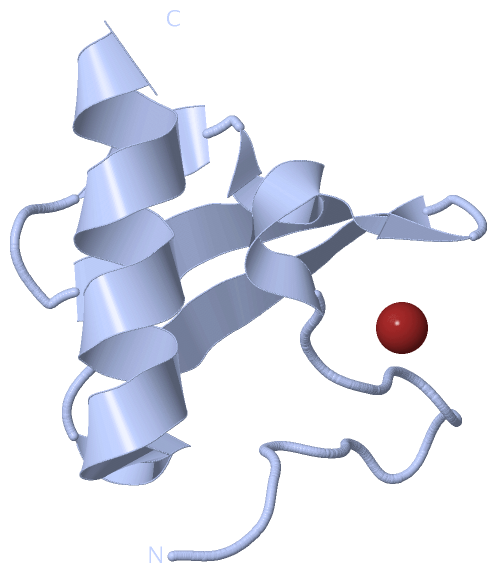
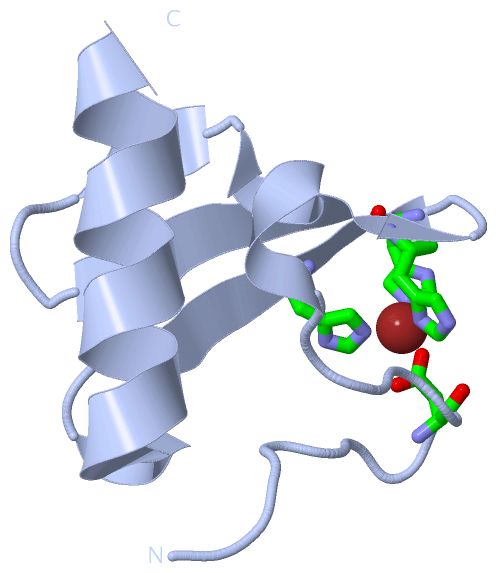
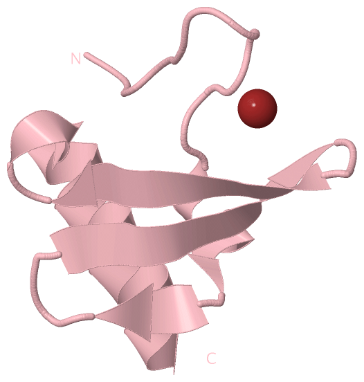
Chain A from PDB  Type:PROTEIN  Length:55
 aligned with A0A0H2UQ89_S | A0A0H2UQ89 from UniProtKB/TrEMBL  Length:802

    Alignment length:55
                                   175       185       195       205       215     
         A0A0H2UQ89_S   166 QGRYTTDDGYIFNASDIIEDTGDAYIVPHGDHYHYIPKNELSASELAAAEAFLSG 220
               SCOP domains d2cs7a1 A:0-54                                          SCOP domains
               CATH domains ------------------------------------------------------- CATH domains
               Pfam domains ------------------------------------------------------- Pfam domains
         Sec.struct. author ............hhhhh.ee...eeeeee..eeeeee.hhhhhhhhhhhhhhhhh Sec.struct. author
                 SAPs(SNPs) ------------------------------------------------------- SAPs(SNPs)
                    PROSITE ------------------------------------------------------- PROSITE
                 Transcript ------------------------------------------------------- Transcript
                 2cs7 A   0 QGRYTTDDGYIFNASDIIEDTGDAYIVPHGDHYHYIPKNELSASELAAAEAFLSG  54
                                     9        19        29        39        49     

Chain B from PDB  Type:PROTEIN  Length:55
 aligned with A0A0H2UQ89_S | A0A0H2UQ89 from UniProtKB/TrEMBL  Length:802

    Alignment length:55
                                   175       185       195       205       215     
         A0A0H2UQ89_S   166 QGRYTTDDGYIFNASDIIEDTGDAYIVPHGDHYHYIPKNELSASELAAAEAFLSG 220
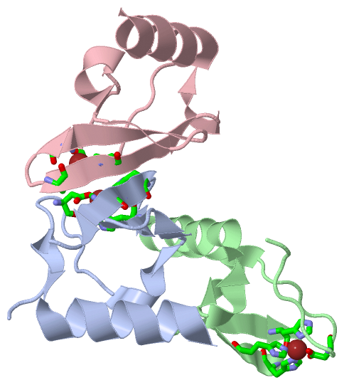
               SCOP domains d2cs7b_ B: Pneumococcal histidine triad protein A, PhtA SCOP domains
               CATH domains ------------------------------------------------------- CATH domains
               Pfam domains ------------------------------------------------------- Pfam domains
         Sec.struct. author ............hhhhh.ee...eeeeee..eeeeee.hhhhhhhhhhhhhhhhh Sec.struct. author
                 SAPs(SNPs) ------------------------------------------------------- SAPs(SNPs)
                    PROSITE ------------------------------------------------------- PROSITE
                 Transcript ------------------------------------------------------- Transcript
                 2cs7 B   0 QGRYTTDDGYIFNASDIIEDTGDAYIVPHGDHYHYIPKNELSASELAAAEAFLSG  54
                                     9        19        29        39        49     

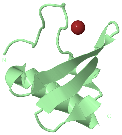
Chain C from PDB  Type:PROTEIN  Length:54
 aligned with A0A0H2UQ89_S | A0A0H2UQ89 from UniProtKB/TrEMBL  Length:802

    Alignment length:54
                                   176       186       196       206       216    
         A0A0H2UQ89_S   167 GRYTTDDGYIFNASDIIEDTGDAYIVPHGDHYHYIPKNELSASELAAAEAFLSG 220
               SCOP domains d2cs7c_ C:                                             SCOP domains
               CATH domains ------------------------------------------------------ CATH domains
               Pfam domains ------------------------------------------------------ Pfam domains
         Sec.struct. author ...........hhhhh.ee...eeeeee..eeeeee.hhhhhhhhhhhhhhhhh Sec.struct. author
                 SAPs(SNPs) ------------------------------------------------------ SAPs(SNPs)
                    PROSITE ------------------------------------------------------ PROSITE
                 Transcript ------------------------------------------------------ Transcript
                 2cs7 C   1 GRYTTDDGYIFNASDIIEDTGDAYIVPHGDHYHYIPKNELSASELAAAEAFLSG  54
                                    10        20        30        40        50    

   Legend:   → Mismatch (orange background)
  - → Gap (green background, '-', border residues have a numbering label)
    → Modified Residue (blue background, lower-case, 'x' indicates undefined single-letter code, labelled with number + name)
  x → Chemical Group (purple background, 'x', labelled with number + name, e.g. ACE or NH2)
  extra numbering lines below/above indicate numbering irregularities and modified residue names etc., number ends below/above '|'

 Classification and Annotation

(-) SCOP Domains  (1, 3)

Asymmetric Unit

(-) CATH Domains  (0, 0)

(no "CATH Domain" information available for 2CS7)

(-) Pfam Domains  (0, 0)

(no "Pfam Domain" information available for 2CS7)

(-) Gene Ontology  (0, 0)

Asymmetric Unit(hide GO term definitions)
    (no "Gene Ontology" information available for 2CS7)

 Visualization

(-) Interactive Views

Asymmetric Unit
  Complete Structure
    Jena3D(integrated viewing of ligand, site, SAP, PROSITE, SCOP information)
    WebMol | AstexViewer[tm]@PDBe
(Java Applets, require no local installation except for Java; loading may be slow)
    STRAP
(Java WebStart application, automatic local installation, requires Java; full application with system access!)
    RasMol
(require local installation)
    Molscript (VRML)
(requires installation of a VRML viewer; select preferred view via VRML and generate a mono or stereo PDF format file)
 
  Ligands, Modified Residues, Ions
    ZN  [ RasMol | Jena3D ]  +environment [ RasMol | Jena3D ]
 
  Sites
    AC1  [ RasMol ]  +environment [ RasMol ]
    AC2  [ RasMol ]  +environment [ RasMol ]
    AC3  [ RasMol ]  +environment [ RasMol ]
 
  Cis Peptide Bonds
(no "Cis Peptide Bonds" information available for 2cs7)
 
Biological Units
  Complete Structure
    Biological Unit 1  [ Jena3D ]
    Biological Unit 2  [ Jena3D ]
    Biological Unit 3  [ Jena3D ]

(-) Still Images

Jmol
  protein: cartoon or spacefill or dots and stick; nucleic acid: cartoon and stick; ligands: spacefill; active site: stick
Molscript
  protein, nucleic acid: cartoon; ligands: spacefill; active site: ball and stick

 Databases and Analysis Tools

(-) Databases

Access by PDB/NDB ID
  2cs7
    Family and Domain InformationProDom | SYSTERS
    General Structural InformationGlycoscienceDB | MMDB | NDB | OCA | PDB | PDBe | PDBj | PDBsum | PDBWiki | PQS | PROTEOPEDIA
    Orientation in MembranesOPM
    Protein SurfaceSURFACE
    Secondary StructureDSSP (structure derived) | HSSP (homology derived)
    Structural GenomicsGeneCensus
    Structural NeighboursCE | VAST
    Structure ClassificationCATH | Dali | SCOP
    Validation and Original DataBMRB Data View | BMRB Restraints Grid | EDS | PROCHECK | RECOORD | WHAT_CHECK
 
Access by UniProt ID/Accession number
  A0A0H2UQ89_S | A0A0H2UQ89
    Comparative Protein Structure ModelsModBase
    Genomic InformationEnsembl
    Protein-protein InteractionDIP
    Sequence, Family and Domain InformationInterPro | Pfam | SMART | UniProtKB/TrEMBL
 
Access by Enzyme Classificator   (EC Number)
  (no 'Enzyme Classificator' available)
    General Enzyme InformationBRENDA | EC-PDB | Enzyme | IntEnz
    PathwayKEGG | MetaCyc
 
Access by Disease Identifier   (MIM ID)
  (no 'MIM ID' available)
    Disease InformationOMIM
 
Access by GenAge ID
  (no 'GenAge ID' available)
    Age Related InformationGenAge

(-) Analysis Tools

Access by PDB/NDB ID
    Domain InformationXDom
    Interatomic Contacts of Structural UnitsCSU
    Ligand-protein ContactsLPC
    Protein CavitiescastP
    Sequence and Secondary StructurePDBCartoon
    Structure AlignmentSTRAP(Java WebStart application, automatic local installation, requires Java; full application with system access!)
    Structure and Sequence BrowserSTING
 
Access by UniProt ID/Accession number
  A0A0H2UQ89_S | A0A0H2UQ89
    Protein Disorder PredictionDisEMBL | FoldIndex | GLOBPLOT (for more information see DisProt)

 Related Entries

(-) Entries Sharing at Least One Protein Chain (UniProt ID)

(no "Entries Sharing at Least One Protein Chain" available for 2CS7)

(-) Related Entries Specified in the PDB File

(no "Related Entries Specified in the PDB File" available for 2CS7)