|
|
|
|
 Description
|
|
Compounds
|
||||||||||||||||||||||||||||||||||||||||||||||||
Chains, Units
Summary Information (see also Sequences/Alignments below) |
Ligands, Modified Residues, Ions (0, 0)| (no "Ligand,Modified Residues,Ions" information available for 2CS4) |
Sites (0, 0)| (no "Site" information available for 2CS4) |
SS Bonds (0, 0)| (no "SS Bond" information available for 2CS4) |
Cis Peptide Bonds (0, 0)| (no "Cis Peptide Bond" information available for 2CS4) |
SAPs(SNPs)/Variants (0, 0)| (no "SAP(SNP)/Variant" information available for 2CS4) |
PROSITE Motifs (1, 1)
NMR Structure (1, 1)
|
||||||||||||||||||||||||
Exons (2, 2)
NMR Structure (2, 2)
|
||||||||||||||||||||||||||||||||||||||||||||||||||||||||||||||||||||||||||||||||||||
Sequences/Alignments
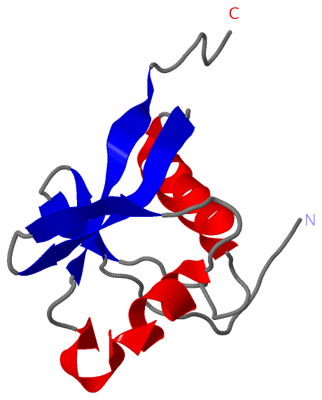
NMR StructureChain A from PDB Type:PROTEIN Length:95 aligned with RASF8_HUMAN | Q8NHQ8 from UniProtKB/Swiss-Prot Length:419 Alignment length:98 1 | 3 13 23 33 43 53 63 73 83 RASF8_HUMAN - -------MELKVWVDGVQRIVCGVTEVTTCQEVVIALAQAIGRTGRYTLIEKWRDTERHLAPHENPIISLNKWGQYASDVQLILRRTGPSLSERPTSD 91 SCOP domains -------d2cs4a1 A:8-91 Ras association domain-containing protein 8 ---- SCOP domains CATH domains -------------------------------------------------------------------------------------------------- CATH domains Pfam domains -------------------------------------------------------------------------------------------------- Pfam domains SAPs(SNPs) -------------------------------------------------------------------------------------------------- SAPs(SNPs) PROSITE -------RA PDB: A:8-89 UniProt: 1-82 --------- PROSITE Transcript 1 (1) -------Exon 1.3 PDB: A:8-42 UniProt: 1-35-------------------------------------------------------- Transcript 1 (1) Transcript 1 (2) -----------------------------------------Exon 1.4 PDB: A:42-95 (gaps) UniProt: 35-331 Transcript 1 (2) 2cs4 A 1 GSSGSSGMELKVWVDGVQRIVCGVTEVTTCQEVVIALAQAIGRTGRYTLIEKWRDTERHLAPHENPIISLNKWGQYASDVQLILRRTGPS---GPSSG 95 10 20 30 40 50 60 70 80 90 | 90 91
|
||||||||||||||||||||
SCOP Domains (1, 1)
NMR Structure
 |
CATH Domains (0, 0)| (no "CATH Domain" information available for 2CS4) |
Pfam Domains (0, 0)| (no "Pfam Domain" information available for 2CS4) |
Gene Ontology (1, 1)|
NMR Structure(hide GO term definitions) Chain A (RASF8_HUMAN | Q8NHQ8)
|
||||||||||||
Interactive Views
|
||||||||||||||||||||||||||||||||||||||||||||||||||||||||||||||||||||||||||||||||||||||||||||||||||||||||||||||||||||
Still Images
|
||||||||||||||||
Databases
|
||||||||||||||||||||||||||||||||||||||||||||||||||||||||||||||||||||||||||||||||||||||||||||||||||||||||||||||||||||||||||||||||||||||||||||||||||||||||||||||||
Analysis Tools
|
|||||||||||||||||||||||||||||||||||||||||||||||||||||||||||||
Entries Sharing at Least One Protein Chain (UniProt ID)
Related Entries Specified in the PDB File
|
|