| molecular function |
|---|
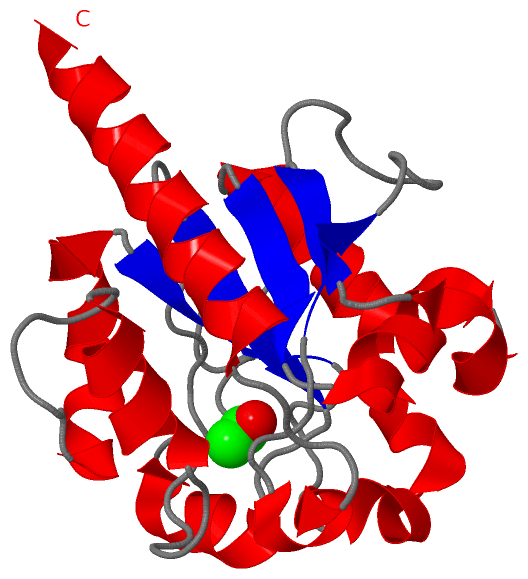
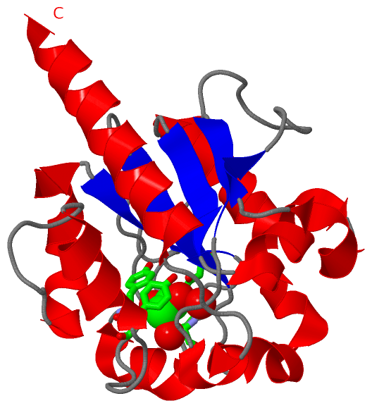
| | GO:0019104 | | DNA N-glycosylase activity | | Catalysis of the removal of damaged bases by cleaving the N-C1' glycosidic bond between the target damaged DNA base and the deoxyribose sugar. The reaction releases a free base and leaves an apurinic/apyrimidinic (AP) site. |
| | GO:0008263 | | pyrimidine-specific mismatch base pair DNA N-glycosylase activity | | Catalysis of the removal of mismatched pyrimidine bases in DNA. Enzymes with this activity recognize and remove pyrimidines present in mismatches by cleaving the N-C1' glycosidic bond between the target damaged DNA base and the deoxyribose sugar. The reaction releases a free base and leaves an apyrimidinic (AP) site. |
| | GO:0004844 | | uracil DNA N-glycosylase activity | | Catalysis of the cleavage of the N-C1' glycosidic bond between the damaged DNA base and the deoxyribose sugar, releasing a free base and leaving an apyrimidinic (AP) site. Enzymes with this activity recognize and remove uracil bases in DNA that result from the deamination of cytosine or the misincorporation of dUTP opposite an adenine. |
| biological process |
|---|
| | GO:0006281 | | DNA repair | | The process of restoring DNA after damage. Genomes are subject to damage by chemical and physical agents in the environment (e.g. UV and ionizing radiations, chemical mutagens, fungal and bacterial toxins, etc.) and by free radicals or alkylating agents endogenously generated in metabolism. DNA is also damaged because of errors during its replication. A variety of different DNA repair pathways have been reported that include direct reversal, base excision repair, nucleotide excision repair, photoreactivation, bypass, double-strand break repair pathway, and mismatch repair pathway. |
| | GO:0006285 | | base-excision repair, AP site formation | | The formation of an AP site, a deoxyribose sugar with a missing base, by DNA glycosylase which recognizes an altered base in DNA and catalyzes its hydrolytic removal. This sugar phosphate is the substrate recognized by the AP endonuclease, which cuts the DNA phosphodiester backbone at the 5' side of the altered site to leave a gap which is subsequently repaired. |
 Description