|
|
|
|
 Description
|
|
Compounds
|
||||||||||||||||||||||||||||||||||||||||||||
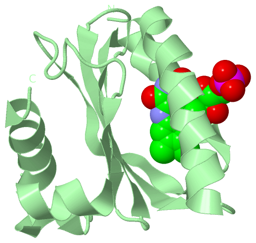
Chains, Units
Summary Information (see also Sequences/Alignments below) |
Ligands, Modified Residues, Ions (1, 2)
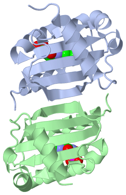
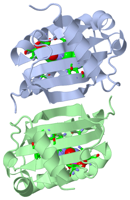
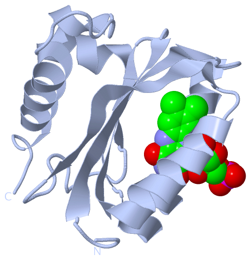
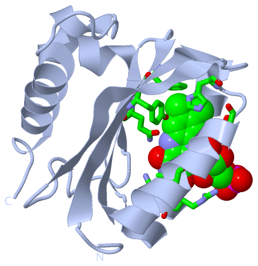
Asymmetric Unit (1, 2)
|
Sites (2, 2)
Asymmetric Unit (2, 2)
|
SS Bonds (0, 0)| (no "SS Bond" information available for 2BYC) |
Cis Peptide Bonds (0, 0)| (no "Cis Peptide Bond" information available for 2BYC) |
SAPs(SNPs)/Variants (0, 0)| (no "SAP(SNP)/Variant" information available for 2BYC) |
PROSITE Motifs (0, 0)| (no "PROSITE Motif" information available for 2BYC) |
Exons (0, 0)| (no "Exon" information available for 2BYC) |
Sequences/Alignments
Asymmetric UnitChain A from PDB Type:PROTEIN Length:137 aligned with Q3IYE4_RHOS4 | Q3IYE4 from UniProtKB/TrEMBL Length:140 Alignment length:137 1 | 9 19 29 39 49 59 69 79 89 99 109 119 129 Q3IYE4_RHOS4 - -MDELVSLTYRSRVRLADPVADIVQIMRASRVRNLRLGITGILLYNGVHFVQTIEGPRSACDELFRLISADPRHQEILAFDLEPITARRFPDWSMRIVSRKELRALAPDLERLDLSGPEDVAELHRTIAASLSKGDA 136 SCOP domains -d2byca1 A:1-136 Blue light receptor BlrB SCOP domains CATH domains ----------------------------------------------------------------------------------------------------------------------------------------- CATH domains Pfam domains ----------------------------------------------------------------------------------------------------------------------------------------- Pfam domains SAPs(SNPs) ----------------------------------------------------------------------------------------------------------------------------------------- SAPs(SNPs) PROSITE ----------------------------------------------------------------------------------------------------------------------------------------- PROSITE Transcript ----------------------------------------------------------------------------------------------------------------------------------------- Transcript 2byc A 0 FMDELVSLTYRSRVRLADPVADIVQIMRASRVRNLRLGITGILLYNGVHFVQTIEGPRSACDELFRLISADPRHQEILAFDLEPITARRFPDWSMRIVSRKELRALAPDLERLDLSGPEDVAELHRTIAASLSRGDA 136 9 19 29 39 49 59 69 79 89 99 109 119 129 Chain B from PDB Type:PROTEIN Length:132 aligned with Q3IYE4_RHOS4 | Q3IYE4 from UniProtKB/TrEMBL Length:140 Alignment length:132 1 | 9 19 29 39 49 59 69 79 89 99 109 119 129 Q3IYE4_RHOS4 - -MDELVSLTYRSRVRLADPVADIVQIMRASRVRNLRLGITGILLYNGVHFVQTIEGPRSACDELFRLISADPRHQEILAFDLEPITARRFPDWSMRIVSRKELRALAPDLERLDLSGPEDVAELHRTIAASL 131 SCOP domains d2bycb_ B: Blue light receptor BlrB SCOP domains CATH domains ------------------------------------------------------------------------------------------------------------------------------------ CATH domains Pfam domains ------------------------------------------------------------------------------------------------------------------------------------ Pfam domains SAPs(SNPs) ------------------------------------------------------------------------------------------------------------------------------------ SAPs(SNPs) PROSITE ------------------------------------------------------------------------------------------------------------------------------------ PROSITE Transcript ------------------------------------------------------------------------------------------------------------------------------------ Transcript 2byc B 500 FMDELVSLTYRSRVRLADPVADIVQIMRASRVRNLRLGITGILLYNGVHFVQTIEGPRSACDELFRLISADPRHQEILAFDLEPITARRFPDWSMRIVSRKELRALAPDLERLDLSGPEDVAELHRTIAASL 631 509 519 529 539 549 559 569 579 589 599 609 619 629
|
||||||||||||||||||||
SCOP Domains (1, 2)
Asymmetric Unit
 |
CATH Domains (0, 0)| (no "CATH Domain" information available for 2BYC) |
Pfam Domains (0, 0)| (no "Pfam Domain" information available for 2BYC) |
Gene Ontology (3, 3)|
Asymmetric Unit(hide GO term definitions) Chain A,B (Q3IYE4_RHOS4 | Q3IYE4)
|
||||||||||||||||||||||||||||||
Interactive Views
|
||||||||||||||||||||||||||||||||||||||||||||||||||||||||||||||||||||||||||||||||||||||||||||||||||||||||||||||||||||||||||||||||||||||||||||||||||||
Still Images
|
||||||||||||||||
Databases
|
||||||||||||||||||||||||||||||||||||||||||||||||||||||||||||||||||||||||||||||||||||||||||||||||||||||||||||||||||||||||||||||||||||||||||||||||||||||||||||||||
Analysis Tools
|
|||||||||||||||||||||||||||||||||||||||||||||||||||||||||||||
Entries Sharing at Least One Protein Chain (UniProt ID)
Related Entries Specified in the PDB File
|
|