Show PDB file:   
         Plain Text   HTML   (compressed file size)
QuickSearch:   
by PDB,NDB,UniProt,PROSITE Code or Search Term(s)  
(-)NMR Structure - manually
(-)NMR Structure - model 1
(-)NMR Structure - all models
collapse expand < >
Image NMR Structure - manually
NMR Structure - manually  (Jmol Viewer)
Image NMR Structure - model 1
NMR Structure - model 1  (Jmol Viewer)
Image NMR Structure - all models
NMR Structure - all models  (Jmol Viewer)

(-) Description

Title :  PERIPHERAL-SUBUNIT BINDING DOMAINS FROM MESOPHILIC, THERMOPHILIC, AND HYPERTHERMOPHILIC BACTERIA FOLD BY ULTRAFAST, APPARENTLY TWO-STATE TRANSITIONS
 
Authors :  N. Ferguson, M. D. Allen
Date :  31 May 05  (Deposition) - 24 May 06  (Release) - 19 Apr 17  (Revision)
Method :  SOLUTION NMR
Resolution :  NOT APPLICABLE
Chains :  NMR Structure  :  A  (20x)
NMR Structure *:  A  (1x)
Keywords :  Acyltransferase, Lipoyl, Transferase (Keyword Search: [Gene Ontology, PubMed, Web (Google))
 
Reference :  N. Ferguson, T. D. Sharpe, P. J. Schartau, S. Sato, M. D. Allen, C. M. Johnson, T. J. Rutherford, A. R. Fersht
Ultra-Fast Barrier-Limited Folding In The Peripheral Subunit-Binding Domain Family.
J. Mol. Biol. V. 353 427 2005
PubMed-ID: 16168437  |  Reference-DOI: 10.1016/J.JMB.2005.08.031

(-) Compounds

Molecule 1 - DIHYDROLIPOYLLYSINE-RESIDUE SUCCINYLTRANSFERASE COMPONENT OF 2-OXOGLUTARATE DEHYDROGENASE COMPLEX
    ChainsA
    EC Number2.3.1.61
    EngineeredYES
    Expression SystemESCHERICHIA COLI
    Expression System StrainC41
    Expression System Taxid562
    Expression System VectorMODIFIED PRSETA
    FragmentRESIDUES 108-152
    MutationYES
    Organism ScientificESCHERICHIA COLI
    Organism Taxid562
    SynonymE3-BINDING DOMAIN, E2

 Structural Features

(-) Chains, Units

  1
NMR Structure (20x)A
NMR Structure * (1x)A

Summary Information (see also Sequences/Alignments below)

(-) Ligands, Modified Residues, Ions  (0, 0)

(no "Ligand,Modified Residues,Ions" information available for 2BTH)

(-) Sites  (0, 0)

(no "Site" information available for 2BTH)

(-) SS Bonds  (0, 0)

(no "SS Bond" information available for 2BTH)

(-) Cis Peptide Bonds  (0, 0)

(no "Cis Peptide Bond" information available for 2BTH)

 Sequence-Structure Mapping

(-) SAPs(SNPs)/Variants  (0, 0)

(no "SAP(SNP)/Variant" information available for 2BTH)

(-) PROSITE Motifs  (1, 1)

NMR Structure (1, 1)
 PROSITEUniProtKBPDB
No.IDACDescriptionIDLocationCountLocation
1PSBDPS51826 Peripheral subunit-binding (PSBD) domain profile.ODO2_ECO57113-150  1A:130-167
ODO2_ECOLI113-150  1A:130-167
NMR Structure * (1, 2)
 PROSITEUniProtKBPDB
No.IDACDescriptionIDLocationCountLocation
1PSBDPS51826 Peripheral subunit-binding (PSBD) domain profile.ODO2_ECO57113-150  1A:130-167
ODO2_ECOLI113-150  1A:130-167

(-) Exons   (0, 0)

(no "Exon" information available for 2BTH)

(-) Sequences/Alignments

NMR Structure
   Reformat: Number of residues per line =  ('0' or empty: single-line sequence representation)
  Number of residues per labelling interval =   
  UniProt sequence: complete  aligned part    
   Show mapping: SCOP domains CATH domains Pfam domains Secondary structure (by author)
SAPs(SNPs) PROSITE motifs Exons
(details for a mapped element are shown in a popup box when the mouse pointer rests over it)
Chain A from PDB  Type:PROTEIN  Length:45
 aligned with ODO2_ECO57 | P0AFG7 from UniProtKB/Swiss-Prot  Length:405

    Alignment length:45
                                   118       128       138       148     
           ODO2_ECO57   109 QNNDALSPAIRRLLAEHNLDASAIKGTGVGGRLTREDVEKHLAKA 153
               SCOP domains d2btha_ A: automated matches                  SCOP domains
               CATH domains --------------------------------------------- CATH domains
               Pfam domains --------------------------------------------- Pfam domains
         Sec.struct. author ......hhhhhhhhhhhh.hhhhh.........hhhhhhhhhhh. Sec.struct. author
                 SAPs(SNPs) --------------------------------------------- SAPs(SNPs)
                    PROSITE ----PSBD  PDB: A:130-167 UniProt: 113-150 --- PROSITE
                 Transcript --------------------------------------------- Transcript
                 2bth A 126 QNNDALSPAIRRLLAEHNLDASAIKGTGVGGRLTREDVEKWLAKA 170
                                   135       145       155       165     

Chain A from PDB  Type:PROTEIN  Length:45
 aligned with ODO2_ECOLI | P0AFG6 from UniProtKB/Swiss-Prot  Length:405

    Alignment length:45
                                   118       128       138       148     
           ODO2_ECOLI   109 QNNDALSPAIRRLLAEHNLDASAIKGTGVGGRLTREDVEKHLAKA 153
               SCOP domains d2btha_ A: automated matches                  SCOP domains
               CATH domains --------------------------------------------- CATH domains
               Pfam domains --------------------------------------------- Pfam domains
         Sec.struct. author ......hhhhhhhhhhhh.hhhhh.........hhhhhhhhhhh. Sec.struct. author
                 SAPs(SNPs) --------------------------------------------- SAPs(SNPs)
                PROSITE (2) ----PSBD  PDB: A:130-167 UniProt: 113-150 --- PROSITE (2)
                 Transcript --------------------------------------------- Transcript
                 2bth A 126 QNNDALSPAIRRLLAEHNLDASAIKGTGVGGRLTREDVEKWLAKA 170
                                   135       145       155       165     

   Legend:   → Mismatch (orange background)
  - → Gap (green background, '-', border residues have a numbering label)
    → Modified Residue (blue background, lower-case, 'x' indicates undefined single-letter code, labelled with number + name)
  x → Chemical Group (purple background, 'x', labelled with number + name, e.g. ACE or NH2)
  extra numbering lines below/above indicate numbering irregularities and modified residue names etc., number ends below/above '|'

 Classification and Annotation

(-) SCOP Domains  (1, 1)

NMR Structure

(-) CATH Domains  (0, 0)

(no "CATH Domain" information available for 2BTH)

(-) Pfam Domains  (0, 0)

(no "Pfam Domain" information available for 2BTH)

(-) Gene Ontology  (10, 17)

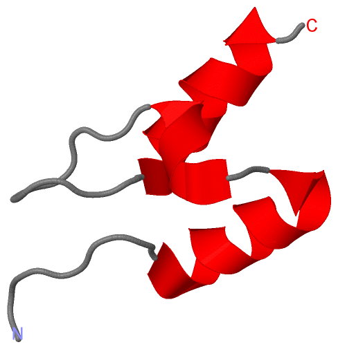
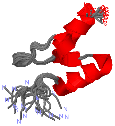
NMR Structure(hide GO term definitions)
Chain A   (ODO2_ECO57 | P0AFG7)
molecular function
    GO:0004149    dihydrolipoyllysine-residue succinyltransferase activity    Catalysis of the reaction: succinyl-CoA + dihydrolipoamide = CoA + S-succinyldihydrolipoamide.
    GO:0016740    transferase activity    Catalysis of the transfer of a group, e.g. a methyl group, glycosyl group, acyl group, phosphorus-containing, or other groups, from one compound (generally regarded as the donor) to another compound (generally regarded as the acceptor). Transferase is the systematic name for any enzyme of EC class 2.
    GO:0016746    transferase activity, transferring acyl groups    Catalysis of the transfer of an acyl group from one compound (donor) to another (acceptor).
biological process
    GO:0033512    L-lysine catabolic process to acetyl-CoA via saccharopine    The chemical reactions and pathways resulting in the breakdown of L-lysine into other compounds, including acetyl-CoA, via the intermediate saccharopine.
    GO:0008152    metabolic process    The chemical reactions and pathways, including anabolism and catabolism, by which living organisms transform chemical substances. Metabolic processes typically transform small molecules, but also include macromolecular processes such as DNA repair and replication, and protein synthesis and degradation.
    GO:0006099    tricarboxylic acid cycle    A nearly universal metabolic pathway in which the acetyl group of acetyl coenzyme A is effectively oxidized to two CO2 and four pairs of electrons are transferred to coenzymes. The acetyl group combines with oxaloacetate to form citrate, which undergoes successive transformations to isocitrate, 2-oxoglutarate, succinyl-CoA, succinate, fumarate, malate, and oxaloacetate again, thus completing the cycle. In eukaryotes the tricarboxylic acid is confined to the mitochondria. See also glyoxylate cycle.
cellular component
    GO:0045252    oxoglutarate dehydrogenase complex    A complex of multiple copies of three enzymatic components: oxoglutarate dehydrogenase (lipoamide) ; EC:1.2.4.2 (E1), dihydrolipoamide S-succinyltransferase ; EC:2.3.1.61 (E2) and dihydrolipoamide dehydrogenase ; EC:1.8.1.4 (E3); catalyzes the overall conversion of 2-oxoglutarate to succinyl-CoA and carbon dioxide (CO2).

Chain A   (ODO2_ECOLI | P0AFG6)
molecular function
    GO:0004149    dihydrolipoyllysine-residue succinyltransferase activity    Catalysis of the reaction: succinyl-CoA + dihydrolipoamide = CoA + S-succinyldihydrolipoamide.
    GO:0031405    lipoic acid binding    Interacting selectively and non-covalently with lipoic acid, 1,2-dithiolane-3-pentanoic acid.
    GO:0005515    protein binding    Interacting selectively and non-covalently with any protein or protein complex (a complex of two or more proteins that may include other nonprotein molecules).
    GO:0016740    transferase activity    Catalysis of the transfer of a group, e.g. a methyl group, glycosyl group, acyl group, phosphorus-containing, or other groups, from one compound (generally regarded as the donor) to another compound (generally regarded as the acceptor). Transferase is the systematic name for any enzyme of EC class 2.
    GO:0016746    transferase activity, transferring acyl groups    Catalysis of the transfer of an acyl group from one compound (donor) to another (acceptor).
biological process
    GO:0033512    L-lysine catabolic process to acetyl-CoA via saccharopine    The chemical reactions and pathways resulting in the breakdown of L-lysine into other compounds, including acetyl-CoA, via the intermediate saccharopine.
    GO:0008152    metabolic process    The chemical reactions and pathways, including anabolism and catabolism, by which living organisms transform chemical substances. Metabolic processes typically transform small molecules, but also include macromolecular processes such as DNA repair and replication, and protein synthesis and degradation.
    GO:0006099    tricarboxylic acid cycle    A nearly universal metabolic pathway in which the acetyl group of acetyl coenzyme A is effectively oxidized to two CO2 and four pairs of electrons are transferred to coenzymes. The acetyl group combines with oxaloacetate to form citrate, which undergoes successive transformations to isocitrate, 2-oxoglutarate, succinyl-CoA, succinate, fumarate, malate, and oxaloacetate again, thus completing the cycle. In eukaryotes the tricarboxylic acid is confined to the mitochondria. See also glyoxylate cycle.
cellular component
    GO:0005829    cytosol    The part of the cytoplasm that does not contain organelles but which does contain other particulate matter, such as protein complexes.
    GO:0045252    oxoglutarate dehydrogenase complex    A complex of multiple copies of three enzymatic components: oxoglutarate dehydrogenase (lipoamide) ; EC:1.2.4.2 (E1), dihydrolipoamide S-succinyltransferase ; EC:2.3.1.61 (E2) and dihydrolipoamide dehydrogenase ; EC:1.8.1.4 (E3); catalyzes the overall conversion of 2-oxoglutarate to succinyl-CoA and carbon dioxide (CO2).

 Visualization

(-) Interactive Views

NMR Structure
  Complete Structure
    Jena3D(integrated viewing of ligand, site, SAP, PROSITE, SCOP information)
    WebMol | AstexViewer[tm]@PDBe
(Java Applets, require no local installation except for Java; loading may be slow)
    STRAP
(Java WebStart application, automatic local installation, requires Java; full application with system access!)
    RasMol
(require local installation)
    Molscript (VRML)
(requires installation of a VRML viewer; select preferred view via VRML and generate a mono or stereo PDF format file)
 
  Ligands, Modified Residues, Ions
(no "Ligands, Modified Residues, Ions" information available for 2bth)
 
  Sites
(no "Sites" information available for 2bth)
 
  Cis Peptide Bonds
(no "Cis Peptide Bonds" information available for 2bth)
 

(-) Still Images

Jmol
  protein: cartoon or spacefill or dots and stick; nucleic acid: cartoon and stick; ligands: spacefill; active site: stick
Molscript
  protein, nucleic acid: cartoon; ligands: spacefill; active site: ball and stick

 Databases and Analysis Tools

(-) Databases

Access by PDB/NDB ID
  2bth
    Family and Domain InformationProDom | SYSTERS
    General Structural InformationGlycoscienceDB | MMDB | NDB | OCA | PDB | PDBe | PDBj | PDBsum | PDBWiki | PQS | PROTEOPEDIA
    Orientation in MembranesOPM
    Protein SurfaceSURFACE
    Secondary StructureDSSP (structure derived) | HSSP (homology derived)
    Structural GenomicsGeneCensus
    Structural NeighboursCE | VAST
    Structure ClassificationCATH | Dali | SCOP
    Validation and Original DataBMRB Data View | BMRB Restraints Grid | EDS | PROCHECK | RECOORD | WHAT_CHECK
 
Access by UniProt ID/Accession number
  ODO2_ECO57 | P0AFG7
    Comparative Protein Structure ModelsModBase
    Genomic InformationEnsembl
    Protein-protein InteractionDIP
    Sequence, Family and Domain InformationInterPro | Pfam | SMART | UniProtKB/SwissProt
  ODO2_ECOLI | P0AFG6
    Comparative Protein Structure ModelsModBase
    Genomic InformationEnsembl
    Protein-protein InteractionDIP
    Sequence, Family and Domain InformationInterPro | Pfam | SMART | UniProtKB/SwissProt
 
Access by Enzyme Classificator   (EC Number)
  2.3.1.61
    General Enzyme InformationBRENDA | EC-PDB | Enzyme | IntEnz
    PathwayKEGG | MetaCyc
 
Access by Disease Identifier   (MIM ID)
  (no 'MIM ID' available)
    Disease InformationOMIM
 
Access by GenAge ID
  (no 'GenAge ID' available)
    Age Related InformationGenAge

(-) Analysis Tools

Access by PDB/NDB ID
    Domain InformationXDom
    Interatomic Contacts of Structural UnitsCSU
    Ligand-protein ContactsLPC
    Protein CavitiescastP
    Sequence and Secondary StructurePDBCartoon
    Structure AlignmentSTRAP(Java WebStart application, automatic local installation, requires Java; full application with system access!)
    Structure and Sequence BrowserSTING
 
Access by UniProt ID/Accession number
  ODO2_ECO57 | P0AFG7
    Protein Disorder PredictionDisEMBL | FoldIndex | GLOBPLOT (for more information see DisProt)
  ODO2_ECOLI | P0AFG6
    Protein Disorder PredictionDisEMBL | FoldIndex | GLOBPLOT (for more information see DisProt)

 Related Entries

(-) Entries Sharing at Least One Protein Chain (UniProt ID)

UniProtKB/Swiss-Prot
        ODO2_ECO57 | P0AFG71bal 1bbl 1c4t 1e2o 1pmr 1scz 2btg 2cyu
        ODO2_ECOLI | P0AFG61bal 1bbl 1c4t 1e2o 1pmr 1scz 1w4h 2btg 2wxc

(-) Related Entries Specified in the PDB File

1bal DIHYDROLIPOAMIDE SUCCINYLTRANSFERASE (E3-BINDING DOMAIN) CORE FROM THE 2-OXOGLUTARATE DEHYDROGENASE MULTIENZYME COMPLEX OF (NMR, 56 SIMULATED ANNEALING STRUCTURES)
1bbl DIHYDROLIPOAMIDE SUCCINYLTRANSFERASE (E3-BINDING DOMAIN) CORE FROM THE 2-OXOGLUTARATE DEHYDROGENASE MULTIENZYME COMPLEX OF ESCHERICHIA COLI (NMR, MINIMIZED AVERAGE STRUCTURE)
1c4t CATALYTIC DOMAIN FROM TRIMERIC DIHYDROLIPOAMIDE SUCCINYLTRANSFERASE
1e2o CATALYTIC DOMAIN FROM DIHYDROLIPOAMIDE SUCCINYLTRANSFERASE
1pmr LIPOYL DOMAIN FROM THE DIHYDROLIPOYL SUCCINYLTRANSFERASE COMPONENT OF THE 2- OXOGLUTARATE DEHYDROGENASE MULTIENZYME COMPLEX OF ESCHERICHIA COLI, NMR, 25 STRUCTURES
1scz IMPROVED STRUCTURAL MODEL FOR THE CATALYTIC DOMAIN OFE.COLI DIHYDROLIPOAMIDE SUCCINYLTRANSFERASE
2btg LIPOYL DOMAIN FROM THE DIHYDROLIPOYL SUCCINYLTRANSFERASE COMPONENT OF THE 2- OXOGLUTARATE DEHYDROGENASE MULTIENZYME COMPLEX OF ESCHERICHIA COLI, NMR, 20 STRUCTURES