| molecular function |
|---|
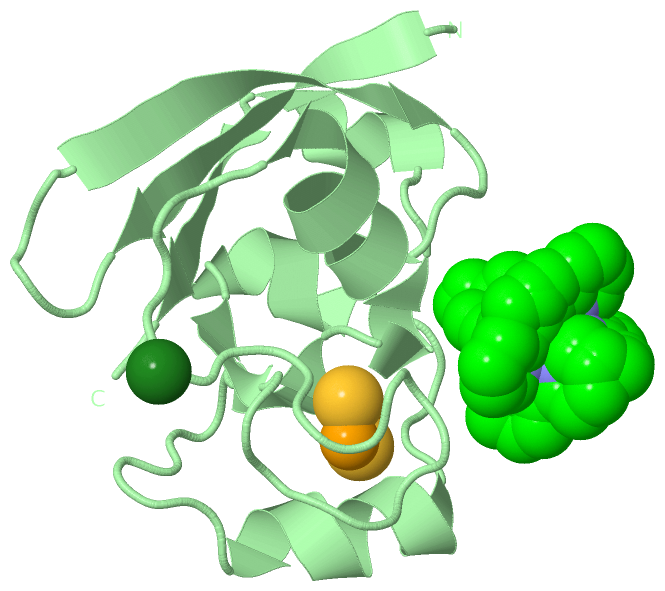
| | GO:0051537 | | 2 iron, 2 sulfur cluster binding | | Interacting selectively and non-covalently with a 2 iron, 2 sulfur (2Fe-2S) cluster; this cluster consists of two iron atoms, with two inorganic sulfur atoms found between the irons and acting as bridging ligands. |
| | GO:0009055 | | electron carrier activity | | Any molecular entity that serves as an electron acceptor and electron donor in an electron transport chain. An electron transport chain is a process in which a series of electron carriers operate together to transfer electrons from donors to any of several different terminal electron acceptors to generate a transmembrane electrochemical gradient. |
| | GO:0051536 | | iron-sulfur cluster binding | | Interacting selectively and non-covalently with an iron-sulfur cluster, a combination of iron and sulfur atoms. |
| | GO:0046872 | | metal ion binding | | Interacting selectively and non-covalently with any metal ion. |
| | GO:0008022 | | protein C-terminus binding | | Interacting selectively and non-covalently with a protein C-terminus, the end of any peptide chain at which the 1-carboxy function of a constituent amino acid is not attached in peptide linkage to another amino-acid residue. |
| | GO:0005515 | | protein binding | | Interacting selectively and non-covalently with any protein or protein complex (a complex of two or more proteins that may include other nonprotein molecules). |
| | GO:0042803 | | protein homodimerization activity | | Interacting selectively and non-covalently with an identical protein to form a homodimer. |
| biological process |
|---|
| | GO:0008203 | | cholesterol metabolic process | | The chemical reactions and pathways involving cholesterol, cholest-5-en-3 beta-ol, the principal sterol of vertebrates and the precursor of many steroids, including bile acids and steroid hormones. It is a component of the plasma membrane lipid bilayer and of plasma lipoproteins and can be found in all animal tissues. |
| | GO:0042446 | | hormone biosynthetic process | | The chemical reactions and pathways resulting in the formation of any hormone, naturally occurring substances secreted by specialized cells that affects the metabolism or behavior of other cells possessing functional receptors for the hormone. |
| | GO:0006629 | | lipid metabolic process | | The chemical reactions and pathways involving lipids, compounds soluble in an organic solvent but not, or sparingly, in an aqueous solvent. Includes fatty acids; neutral fats, other fatty-acid esters, and soaps; long-chain (fatty) alcohols and waxes; sphingoids and other long-chain bases; glycolipids, phospholipids and sphingolipids; and carotenes, polyprenols, sterols, terpenes and other isoprenoids. |
| | GO:0055114 | | oxidation-reduction process | | A metabolic process that results in the removal or addition of one or more electrons to or from a substance, with or without the concomitant removal or addition of a proton or protons. |
| | GO:0006694 | | steroid biosynthetic process | | The chemical reactions and pathways resulting in the formation of steroids, compounds with a 1,2,cyclopentanoperhydrophenanthrene nucleus; includes de novo formation and steroid interconversion by modification. |
| | GO:0008202 | | steroid metabolic process | | The chemical reactions and pathways involving steroids, compounds with a 1,2,cyclopentanoperhydrophenanthrene nucleus. |
| cellular component |
|---|
| | GO:0005759 | | mitochondrial matrix | | The gel-like material, with considerable fine structure, that lies in the matrix space, or lumen, of a mitochondrion. It contains the enzymes of the tricarboxylic acid cycle and, in some organisms, the enzymes concerned with fatty acid oxidation. |
| | GO:0005739 | | mitochondrion | | A semiautonomous, self replicating organelle that occurs in varying numbers, shapes, and sizes in the cytoplasm of virtually all eukaryotic cells. It is notably the site of tissue respiration. |
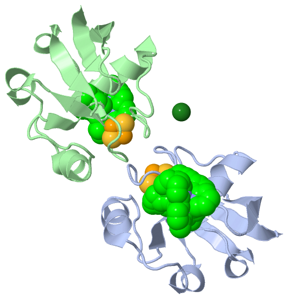
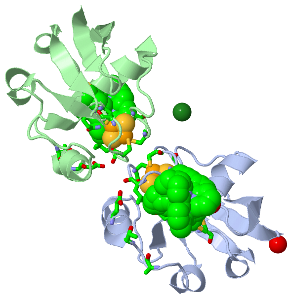
 Description
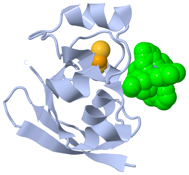
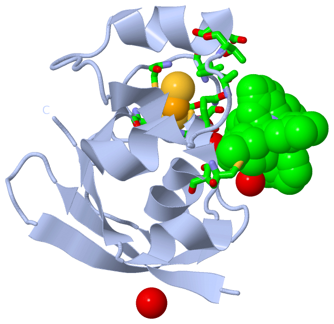