|
|
|
|
 Description
|
|
Compounds
|
||||||||||||||||||||||||||||||||||||||||||||||||||||
Chains, Units
Summary Information (see also Sequences/Alignments below) |
Ligands, Modified Residues, Ions (4, 14)| Asymmetric/Biological Unit (4, 14) |
Sites (3, 3)
Asymmetric Unit (3, 3)
|
SS Bonds (0, 0)| (no "SS Bond" information available for 2BE3) |
Cis Peptide Bonds (0, 0)| (no "Cis Peptide Bond" information available for 2BE3) |
SAPs(SNPs)/Variants (0, 0)| (no "SAP(SNP)/Variant" information available for 2BE3) |
PROSITE Motifs (0, 0)| (no "PROSITE Motif" information available for 2BE3) |
Exons (0, 0)| (no "Exon" information available for 2BE3) |
Sequences/Alignments
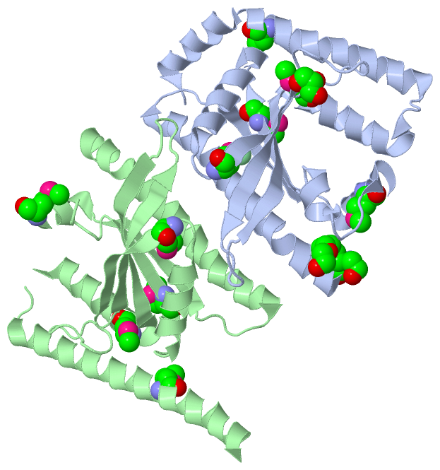
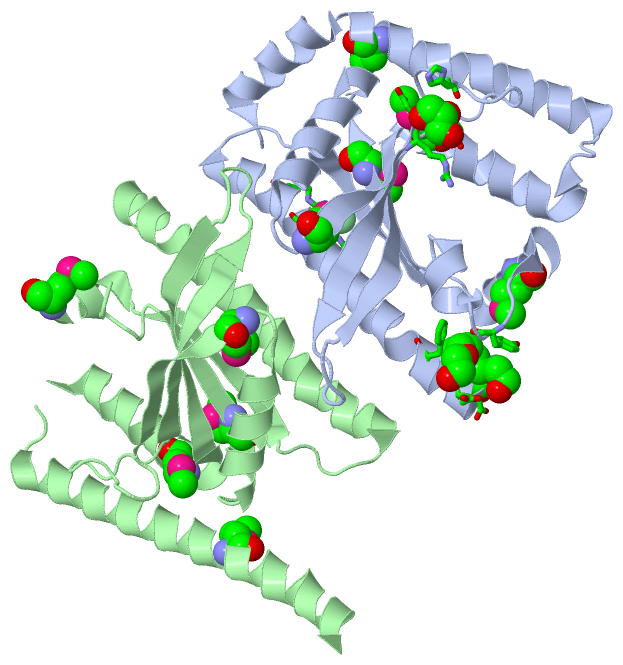
Asymmetric/Biological UnitChain A from PDB Type:PROTEIN Length:203 aligned with A0A0H2UPY1_S | A0A0H2UPY1 from UniProtKB/TrEMBL Length:223 Alignment length:203 10 20 30 40 50 60 70 80 90 100 110 120 130 140 150 160 170 180 190 200 A0A0H2UPY1_S 1 MTLEWEEFLDPYIQAVGELKIKLRGIRKQYRKQNKHSPIEFVTGRVKPIESIKEKMARRGITYATLEHDLQDIAGLRVMVQFVDDVKEVVDILHKRQDMRIIQERDYITHRKASGYRSYHVVVEYTVDTINGAKTILAEIQIRTLAMNFWATIEHSLNYKYQGDFPDEIKKRLEITARIAHQLDEEMGEIRDDIQEAQALFDP 203 SCOP domains d2be3a1 A:1-203 Putative GTP pyrophosphokinase SP1097 SCOP domains CATH domains -2be3A01 A:2-146 Beta Polymerase, domain 2 -2be3A02 A:148-203 Nucleotidyltransferase CATH domains Pfam domains ----------------------------------------------------------------------------------------------------------------------------------------------------------------------------------------------------------- Pfam domains SAPs(SNPs) ----------------------------------------------------------------------------------------------------------------------------------------------------------------------------------------------------------- SAPs(SNPs) PROSITE ----------------------------------------------------------------------------------------------------------------------------------------------------------------------------------------------------------- PROSITE Transcript ----------------------------------------------------------------------------------------------------------------------------------------------------------------------------------------------------------- Transcript 2be3 A 1 mTLEWEEFLDPYIQAVGELKIKLRGIRKQYRKQNKHSPIEFVTGRVKPIESIKEKmARRGITYATLEHDLQDIAGLRVmVQFVDDVKEVVDILHKRQDmRIIQERDYITHRKASGYRSYHVVVEYTVDTINGAKTILAEIQIRTLAmNFWATIEHSLNYKYQGDFPDEIKKRLEITARIAHQLDEEmGEIRDDIQEAQALFDP 203 | 10 20 30 40 50 | 60 70 80 90 100 110 120 130 140 |150 160 170 180 |190 200 | 56-MSE 79-MSE 99-MSE 147-MSE 187-MSE 1-MSE Chain B from PDB Type:PROTEIN Length:189 aligned with A0A0H2UPY1_S | A0A0H2UPY1 from UniProtKB/TrEMBL Length:223 Alignment length:200 11 21 31 41 51 61 71 81 91 101 111 121 131 141 151 161 171 181 191 201 A0A0H2UPY1_S 2 TLEWEEFLDPYIQAVGELKIKLRGIRKQYRKQNKHSPIEFVTGRVKPIESIKEKMARRGITYATLEHDLQDIAGLRVMVQFVDDVKEVVDILHKRQDMRIIQERDYITHRKASGYRSYHVVVEYTVDTINGAKTILAEIQIRTLAMNFWATIEHSLNYKYQGDFPDEIKKRLEITARIAHQLDEEMGEIRDDIQEAQALF 201 SCOP domains d2be3b_ B: Putative GTP pyrophosphokinase SP1097 SCOP domains CATH domains 2be3B01 B:2-146 Beta Polymerase, domain 2 -2be3B02 B:148-2 01 Nucleotidyltransferase CATH domains Pfam domains -------------------------------------------------------------------------------------------------------------------------------------------------------------------------------------------------------- Pfam domains SAPs(SNPs) -------------------------------------------------------------------------------------------------------------------------------------------------------------------------------------------------------- SAPs(SNPs) PROSITE -------------------------------------------------------------------------------------------------------------------------------------------------------------------------------------------------------- PROSITE Transcript -------------------------------------------------------------------------------------------------------------------------------------------------------------------------------------------------------- Transcript 2be3 B 2 TLEWEEFLDPYIQAVGELKIKLRGIRKQYRKQNKHSPIEFVTGRVKPIESIKEKmA----------HDLQDIAGLRVmVQFVDDVKEVVDILHKRQDmRIIQERDYITHRKASGYRSYHVVVEYTVDTINGAKTILAEIQIRTLAmNFWATIEHSLNYKYQ-DFPDEIKKRLEITARIAHQLDEEmGEIRDDIQEAQALF 201 11 21 31 41 51 || - | 71 |81 91 101 111 121 131 141 | 151 161| | 171 181 | 191 201 56-MSE 68 79-MSE 99-MSE 147-MSE 162 | 187-MSE 57 164
|
||||||||||||||||||||
SCOP Domains (1, 2)
Asymmetric/Biological Unit
 |
CATH Domains (2, 4)
Asymmetric/Biological Unit
|
Pfam Domains (0, 0)| (no "Pfam Domain" information available for 2BE3) |
Gene Ontology (0, 0)|
Asymmetric/Biological Unit(hide GO term definitions)
(no "Gene Ontology" information available for 2BE3)
|
Interactive Views
|
|||||||||||||||||||||||||||||||||||||||||||||||||||||||||||||||||||||||||||||||||||||||||||||||||||||||||||||||||||||||||||||||||||||||||||||||||||||||||
Still Images
|
||||||||||||||||
Databases
|
||||||||||||||||||||||||||||||||||||||||||||||||||||||||||||||||||||||||||||||||||||||||||||||||||||||||||||||||||||||||||||||||||||||||||||||||||||||||||||||||
Analysis Tools
|
|||||||||||||||||||||||||||||||||||||||||||||||||||||||||||||
Entries Sharing at Least One Protein Chain (UniProt ID)
Related Entries Specified in the PDB File
|
|