Show PDB file:   
         Plain Text   HTML   (compressed file size)
QuickSearch:   
by PDB,NDB,UniProt,PROSITE Code or Search Term(s)  
(-)NMR Structure - model 1
(-)NMR Structure - model 1, sites
(-)NMR Structure - all models
collapse expand < >
Image NMR Structure - model 1
NMR Structure - model 1  (Jmol Viewer)
Image NMR Structure - model 1, sites
NMR Structure - model 1, sites  (Jmol Viewer)
Image NMR Structure - all models
NMR Structure - all models  (Jmol Viewer)

(-) Description

Title :  THREE-DIMENSIONAL STRUCTURE OF SOYBEAN TRYPSIN(SLASH)CHYMOTRYPSIN BOWMAN-BIRK INHIBITOR IN SOLUTION
 
Authors :  M. H. Werner, D. E. Wemmer
Date :  19 Sep 91  (Deposition) - 31 Oct 93  (Release) - 25 Sep 13  (Revision)
Method :  SOLUTION NMR
Resolution :  NOT APPLICABLE
Chains :  NMR Structure  :  A  (16x)
NMR Structure *:  A  (1x)
Keywords :  Serine Protease Inhibitor (Keyword Search: [Gene Ontology, PubMed, Web (Google))
 
Reference :  M. H. Werner, D. E. Wemmer
Three-Dimensional Structure Of Soybean Trypsin/Chymotrypsin Bowman-Birk Inhibitor In Solution.
Biochemistry V. 31 999 1992
PubMed-ID: 1734975  |  Reference-DOI: 10.1021/BI00119A008
(for further references see the PDB file header)

(-) Compounds

Molecule 1 - TRYPSIN/CHYMOTRYPSIN BOWMAN-BIRK INHIBITOR
    ChainsA
    EngineeredYES
    Organism CommonSOYBEAN
    Organism ScientificGLYCINE MAX
    Organism Taxid3847

 Structural Features

(-) Chains, Units

  1
NMR Structure (16x)A
NMR Structure * (1x)A

Summary Information (see also Sequences/Alignments below)

(-) Ligands, Modified Residues, Ions  (0, 0)

(no "Ligand,Modified Residues,Ions" information available for 2BBI)
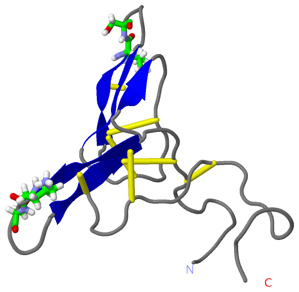
(-) Sites  (2, 2)

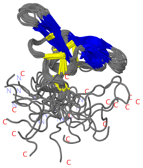
NMR Structure (2, 2)
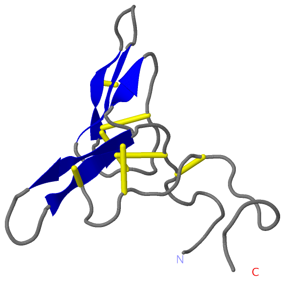
No.NameEvidenceResiduesDescription
1C1UNKNOWNLEU A:43 , SER A:44NULL
2T1UNKNOWNLYS A:16 , SER A:17NULL

(-) SS Bonds  (7, 7)

NMR Structure
No.Residues
1A:8 -A:62
2A:9 -A:24
3A:12 -A:58
4A:14 -A:22
5A:32 -A:39
6A:36 -A:51
7A:41 -A:49

(-) Cis Peptide Bonds  (2, 32)

NMR Structure
No.ModelResidues
11, 2, 3, 4, 5, 6, 7, 8, 9, 10, 11, 12, 13, 14, 15, 16Asn A:18 -Pro A:19
21, 2, 3, 4, 5, 6, 7, 8, 9, 10, 11, 12, 13, 14, 15, 16Tyr A:45 -Pro A:46

 Sequence-Structure Mapping

(-) SAPs(SNPs)/Variants  (0, 0)

(no "SAP(SNP)/Variant" information available for 2BBI)

(-) PROSITE Motifs  (1, 1)

NMR Structure (1, 1)
 PROSITEUniProtKBPDB
No.IDACDescriptionIDLocationCountLocation
1BOWMAN_BIRKPS00281 Bowman-Birk serine protease inhibitors family signature.IBB1_SOYBN63-78  1A:24-39
NMR Structure * (1, 1)
 PROSITEUniProtKBPDB
No.IDACDescriptionIDLocationCountLocation
1BOWMAN_BIRKPS00281 Bowman-Birk serine protease inhibitors family signature.IBB1_SOYBN63-78  1A:24-39

(-) Exons   (0, 0)

(no "Exon" information available for 2BBI)

(-) Sequences/Alignments

NMR Structure
   Reformat: Number of residues per line =  ('0' or empty: single-line sequence representation)
  Number of residues per labelling interval =   
  UniProt sequence: complete  aligned part    
   Show mapping: SCOP domains CATH domains Pfam domains Secondary structure (by author)
SAPs(SNPs) PROSITE motifs Exons
(details for a mapped element are shown in a popup box when the mouse pointer rests over it)
Chain A from PDB  Type:PROTEIN  Length:71
 aligned with IBB1_SOYBN | P01055 from UniProtKB/Swiss-Prot  Length:110

    Alignment length:71
                                    49        59        69        79        89        99       109 
           IBB1_SOYBN    40 DDESSKPCCDQCACTKSNPPQCRCSDMRLNSCHSACKSCICALSYPAQCFCVDITDFCYEPCKPSEDDKEN 110
               SCOP domains d2bbia_ A: Bowman-Birk inhibitor, BBI                                   SCOP domains
               CATH domains 2bbiA00 A:1-71 Cysteine Protease (Bromelain) Inhibitor, subunit H       CATH domains
               Pfam domains ----------------------------------------------------------------------- Pfam domains
         Sec.struct. author ...........eeee.....eeee..ee..........eeee.....eeee.................... Sec.struct. author
                 SAPs(SNPs) ----------------------------------------------------------------------- SAPs(SNPs)
                    PROSITE -----------------------BOWMAN_BIRK     -------------------------------- PROSITE
                 Transcript ----------------------------------------------------------------------- Transcript
                 2bbi A   1 DDESSKPCCDQCACTKSNPPQCRCSDMRLNSCHSACKSCICALSYPAQCFCVDITDFCYEPCKPSEDDKEN  71
                                    10        20        30        40        50        60        70 

   Legend:   → Mismatch (orange background)
  - → Gap (green background, '-', border residues have a numbering label)
    → Modified Residue (blue background, lower-case, 'x' indicates undefined single-letter code, labelled with number + name)
  x → Chemical Group (purple background, 'x', labelled with number + name, e.g. ACE or NH2)
  extra numbering lines below/above indicate numbering irregularities and modified residue names etc., number ends below/above '|'

 Classification and Annotation

(-) SCOP Domains  (1, 1)

NMR Structure

(-) CATH Domains  (1, 1)

NMR Structure
(-)
Class: Mainly Beta (13760)

(-) Pfam Domains  (0, 0)

(no "Pfam Domain" information available for 2BBI)

(-) Gene Ontology  (5, 5)

NMR Structure(hide GO term definitions)
Chain A   (IBB1_SOYBN | P01055)
molecular function
    GO:0030414    peptidase inhibitor activity    Stops, prevents or reduces the activity of a peptidase, any enzyme that catalyzes the hydrolysis peptide bonds.
    GO:0004867    serine-type endopeptidase inhibitor activity    Stops, prevents or reduces the activity of serine-type endopeptidases, enzymes that catalyze the hydrolysis of nonterminal peptide bonds in a polypeptide chain; a serine residue (and a histidine residue) are at the active center of the enzyme.
biological process
    GO:0010951    negative regulation of endopeptidase activity    Any process that decreases the frequency, rate or extent of endopeptidase activity, the endohydrolysis of peptide bonds within proteins.
    GO:0010466    negative regulation of peptidase activity    Any process that stops or reduces the rate of peptidase activity, the hydrolysis of peptide bonds within proteins.
cellular component
    GO:0005576    extracellular region    The space external to the outermost structure of a cell. For cells without external protective or external encapsulating structures this refers to space outside of the plasma membrane. This term covers the host cell environment outside an intracellular parasite.

 Visualization

(-) Interactive Views

NMR Structure
  Complete Structure
    Jena3D(integrated viewing of ligand, site, SAP, PROSITE, SCOP information)
    WebMol | AstexViewer[tm]@PDBe
(Java Applets, require no local installation except for Java; loading may be slow)
    STRAP
(Java WebStart application, automatic local installation, requires Java; full application with system access!)
    RasMol
(require local installation)
    Molscript (VRML)
(requires installation of a VRML viewer; select preferred view via VRML and generate a mono or stereo PDF format file)
 
  Ligands, Modified Residues, Ions
(no "Ligands, Modified Residues, Ions" information available for 2bbi)
 
  Sites
    C1  [ RasMol ]  +environment [ RasMol ]
    T1  [ RasMol ]  +environment [ RasMol ]
 
  Cis Peptide Bonds
    Asn A:18 - Pro A:19   [ RasMol ]  
    Tyr A:45 - Pro A:46   [ RasMol ]  
 

(-) Still Images

Jmol
  protein: cartoon or spacefill or dots and stick; nucleic acid: cartoon and stick; ligands: spacefill; active site: stick
Molscript
  protein, nucleic acid: cartoon; ligands: spacefill; active site: ball and stick

 Databases and Analysis Tools

(-) Databases

Access by PDB/NDB ID
  2bbi
    Family and Domain InformationProDom | SYSTERS
    General Structural InformationGlycoscienceDB | MMDB | NDB | OCA | PDB | PDBe | PDBj | PDBsum | PDBWiki | PQS | PROTEOPEDIA
    Orientation in MembranesOPM
    Protein SurfaceSURFACE
    Secondary StructureDSSP (structure derived) | HSSP (homology derived)
    Structural GenomicsGeneCensus
    Structural NeighboursCE | VAST
    Structure ClassificationCATH | Dali | SCOP
    Validation and Original DataBMRB Data View | BMRB Restraints Grid | EDS | PROCHECK | RECOORD | WHAT_CHECK
 
Access by UniProt ID/Accession number
  IBB1_SOYBN | P01055
    Comparative Protein Structure ModelsModBase
    Genomic InformationEnsembl
    Protein-protein InteractionDIP
    Sequence, Family and Domain InformationInterPro | Pfam | SMART | UniProtKB/SwissProt
 
Access by Enzyme Classificator   (EC Number)
  (no 'Enzyme Classificator' available)
    General Enzyme InformationBRENDA | EC-PDB | Enzyme | IntEnz
    PathwayKEGG | MetaCyc
 
Access by Disease Identifier   (MIM ID)
  (no 'MIM ID' available)
    Disease InformationOMIM
 
Access by GenAge ID
  (no 'GenAge ID' available)
    Age Related InformationGenAge

(-) Analysis Tools

Access by PDB/NDB ID
    Domain InformationXDom
    Interatomic Contacts of Structural UnitsCSU
    Ligand-protein ContactsLPC
    Protein CavitiescastP
    Sequence and Secondary StructurePDBCartoon
    Structure AlignmentSTRAP(Java WebStart application, automatic local installation, requires Java; full application with system access!)
    Structure and Sequence BrowserSTING
 
Access by UniProt ID/Accession number
  IBB1_SOYBN | P01055
    Protein Disorder PredictionDisEMBL | FoldIndex | GLOBPLOT (for more information see DisProt)

 Related Entries

(-) Entries Sharing at Least One Protein Chain (UniProt ID)

UniProtKB/Swiss-Prot
        IBB1_SOYBN | P010551bbi 1d6r 1k9b 5j4q 5j4s

(-) Related Entries Specified in the PDB File

1bbi