|
|
|
|
 Description
|
|
Compounds
|
||||||||||||||||||||||||||||||||||||||||||||||||||||||||
Chains, Units
Summary Information (see also Sequences/Alignments below) |
Ligands, Modified Residues, Ions (2, 3)| Asymmetric/Biological Unit (2, 3) |
Sites (3, 3)
Asymmetric Unit (3, 3)
|
SS Bonds (2, 2)
Asymmetric/Biological Unit
|
||||||||||||
Cis Peptide Bonds (3, 3)
Asymmetric/Biological Unit
|
||||||||||||||||
SAPs(SNPs)/Variants (0, 0)| (no "SAP(SNP)/Variant" information available for 2B5E) |
PROSITE Motifs (2, 3)
Asymmetric/Biological Unit (2, 3)
|
||||||||||||||||||||||||||||||||
Exons (1, 1)| Asymmetric/Biological Unit (1, 1) |
Sequences/Alignments
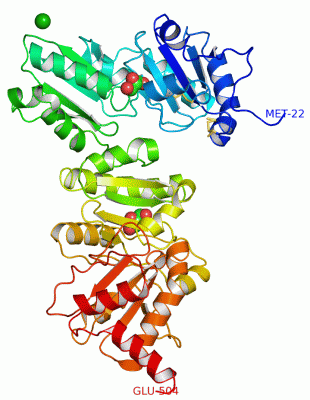
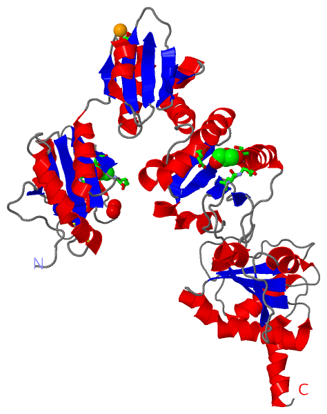
Asymmetric/Biological UnitChain A from PDB Type:PROTEIN Length:483 aligned with PDI_YEAST | P17967 from UniProtKB/Swiss-Prot Length:522 Alignment length:483 31 41 51 61 71 81 91 101 111 121 131 141 151 161 171 181 191 201 211 221 231 241 251 261 271 281 291 301 311 321 331 341 351 361 371 381 391 401 411 421 431 441 451 461 471 481 491 501 PDI_YEAST 22 AQQEAVAPEDSAVVKLATDSFNEYIQSHDLVLAEFFAPWCGHCKNMAPEYVKAAETLVEKNITLAQIDCTENQDLCMEHNIPGFPSLKIFKNSDVNNSIDYEGPRTAEAIVQFMIKQSQPAVAVVADLPAYLANETFVTPVIVQSGKIDADFNATFYSMANKHFNDYDFVSAENADDDFKLSIYLPSAMDEPVVYNGKKADIADADVFEKWLQVEALPYFGEIDGSVFAQYVESGLPLGYLFYNDEEELEEYKPLFTELAKKNRGLMNFVSIDARKFGRHAGNLNMKEQFPLFAIHDMTEDLKYGLPQLSEEAFDELSDKIVLESKAIESLVKDFLKGDASPIVKSQEIFENQDSSVFQLVGKNHDEIVNDPKKDVLVLYYAPWCGHCKRLAPTYQELADTYANATSDVLIAKLDHTENDVRGVVIEGYPTIVLYPGGKKSESVVYQGSRSLDSLFDFIKENGHFDVDGKALYEEAQEKAAEE 504 SCOP domains -d2b5ea4 A:23-141 Protein disulfide isomerase, PDI d2b5ea2 A:142-239 Protein disulfide isomerase, PDI d2b5ea3 A:240-364 Protein disulfide isomerase, PDI d2b5ea1 A:365-504 Protein disulfide isomerase, PDI SCOP domains CATH domains 2b5eA01 A:22-140 Glutaredoxin --------------------------------------------------------------------------------------------------------------------------------------------------------------------------------------------------------------------------------2b5eA04 A:365-504 Glutaredoxin CATH domains Pfam domains --------------------------------------------------------------------------------------------------------------------------------------------------------------------------------------------------------------------------------------------------------------------------------------------------------------------------------------------------------------------------------------------------------------------------------------------------------------------------------------------------- Pfam domains SAPs(SNPs) --------------------------------------------------------------------------------------------------------------------------------------------------------------------------------------------------------------------------------------------------------------------------------------------------------------------------------------------------------------------------------------------------------------------------------------------------------------------------------------------------- SAPs(SNPs) PROSITE (1) THIOREDOXIN_2 PDB: - UniProt: 15-141 ----------------------------------------------------------------------------------------------------------------------------------------------------------------------------------------------------------------------THIOREDOXIN_2 PDB: A:356-485 UniProt: 356-485 ------------------- PROSITE (1) PROSITE (2) -------------------------------THIOREDOXIN_1 --------------------------------------------------------------------------------------------------------------------------------------------------------------------------------------------------------------------------------------------------------------------------------------------------------------------------------------THIOREDOXIN_1 ---------------------------------------------------------------------------------------- PROSITE (2) Transcript 1 Exon 1.1 PDB: A:22-504 UniProt: 1-522 [INCOMPLETE] Transcript 1 2b5e A 22 MQQEAVAPEDSAVVKLATDSFNEYIQSHDLVLAEFFAPWCGHCKNMAPEYVKAAETLVEKNITLAQIDCTENQDLCMEHNIPGFPSLKIFKNSDVNNSIDYEGPRTAEAIVQFMIKQSQPAVAVVADLPAYLANETFVTPVIVQSGKIDADFNATFYSMANKHFNDYDFVSAENADDDFKLSIYLPSAMDEPVVYNGKKADIADADVFEKWLQVEALPYFGEIDGSVFAQYVESGLPLGYLFYNDEEELEEYKPLFTELAKKNRGLMNFVSIDARKFGRHAGNLNMKEQFPLFAIHDMTEDLKYGLPQLSEEAFDELSDKIVLESKAIESLVKDFLKGDASPIVKSQEIFENQDSSVFQLVGKNHDEIVNDPKKDVLVLYYAPWCGHCKRLAPTYQELADTYANATSDVLIAKLDHTENDVRGVVIEGYPTIVLYPGGKKSESVVYQGSRSLDSLFDFIKENGHFDVDGKALYEEAQEKAAEE 504 31 41 51 61 71 81 91 101 111 121 131 141 151 161 171 181 191 201 211 221 231 241 251 261 271 281 291 301 311 321 331 341 351 361 371 381 391 401 411 421 431 441 451 461 471 481 491 501
|
||||||||||||||||||||
SCOP Domains (1, 4)
Asymmetric/Biological Unit
 |
CATH Domains (1, 2)
Asymmetric/Biological Unit
|
Pfam Domains (0, 0)| (no "Pfam Domain" information available for 2B5E) |
Gene Ontology (11, 11)|
Asymmetric/Biological Unit(hide GO term definitions) Chain A (PDI_YEAST | P17967)
|
||||||||||||||||||||||||||||||||||||||||||||||||||||||||||||||||||||||||||||||||||||
Interactive Views
|
||||||||||||||||||||||||||||||||||||||||||||||||||||||||||||||||||||||||||||||||||||||||||||||||||||||||||||||||||||||||||||||||||||||||||||||||||||||||||
Still Images
|
||||||||||||||||
Databases
|
||||||||||||||||||||||||||||||||||||||||||||||||||||||||||||||||||||||||||||||||||||||||||||||||||||||||||||||||||||||||||||||||||||||||||||||||||||||||||||||||
Analysis Tools
|
|||||||||||||||||||||||||||||||||||||||||||||||||||||||||||||
Entries Sharing at Least One Protein Chain (UniProt ID)
Related Entries Specified in the PDB File
|
|