|
|
|
|
 Description
|
|
Compounds
|
||||||||||||||||||||||||||||||||||||||||||||||||
Chains, Units
Summary Information (see also Sequences/Alignments below) |
Ligands, Modified Residues, Ions (4, 5)
Asymmetric Unit (4, 5)
|
Sites (3, 3)
Asymmetric Unit (3, 3)
|
SS Bonds (0, 0)| (no "SS Bond" information available for 2B0V) |
Cis Peptide Bonds (0, 0)| (no "Cis Peptide Bond" information available for 2B0V) |
SAPs(SNPs)/Variants (0, 0)| (no "SAP(SNP)/Variant" information available for 2B0V) |
PROSITE Motifs (0, 0)| (no "PROSITE Motif" information available for 2B0V) |
Exons (0, 0)| (no "Exon" information available for 2B0V) |
Sequences/Alignments
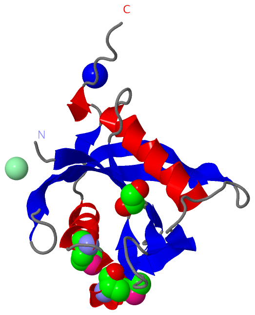
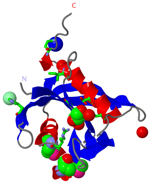
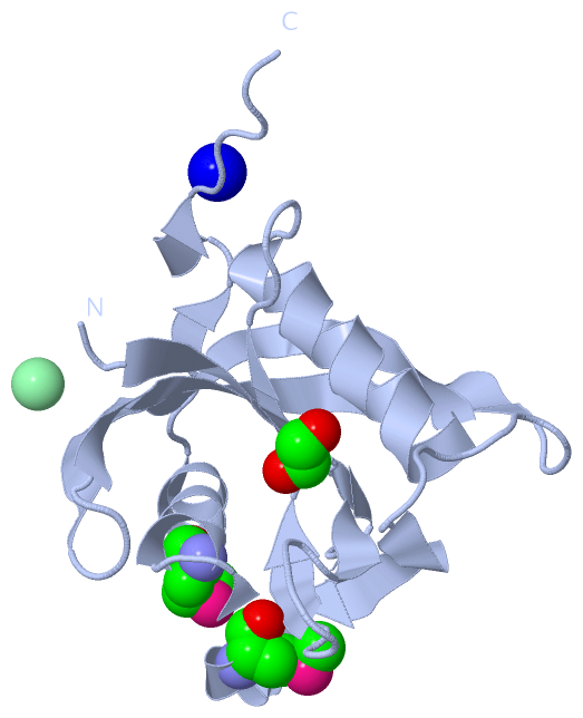
Asymmetric UnitChain A from PDB Type:PROTEIN Length:148 aligned with Q82XR9_NITEU | Q82XR9 from UniProtKB/TrEMBL Length:149 Alignment length:148 149 13 23 33 43 53 63 73 83 93 103 113 123 133 143 | Q82XR9_NITEU 4 KPNVTVAAVIEQDDKYLLVEEIPRGTAIKLNQPAGHLEPGESIIQACSREVLEETGHSFLPEVLTGIYHWTCASNGTTYLRFTFSGQVVSFDPDRKLDTGIVRAAWFSIDEIRAKQAMHRTPLVMQCIEDYHAGKRYPLDILQYYD-- - SCOP domains d2b0va1 A:4-149 Hypothetical protein NE0184 -- SCOP domains CATH domains 2b0vA00 A:4-151 Nucleoside Triphosphate Pyrophosphohydrolase CATH domains Pfam domains ---------------------------------------------------------------------------------------------------------------------------------------------------- Pfam domains SAPs(SNPs) ---------------------------------------------------------------------------------------------------------------------------------------------------- SAPs(SNPs) PROSITE ---------------------------------------------------------------------------------------------------------------------------------------------------- PROSITE Transcript ---------------------------------------------------------------------------------------------------------------------------------------------------- Transcript 2b0v A 4 KPNVTVAAVIEQDDKYLLVEEIPRGTAIKLNQPAGHLEPGESIIQACSREVLEETGHSFLPEVLTGIYHWTCASNGTTYLRFTFSGQVVSFDPDRKLDTGIVRAAWFSIDEIRAKQAmHRTPLVmQCIEDYHAGKRYPLDILQYYDGS 151 13 23 33 43 53 63 73 83 93 103 113 123 | 133 143 121-MSE | 128-MSE
|
||||||||||||||||||||
SCOP Domains (1, 1)
Asymmetric Unit

|
CATH Domains (1, 1)
Asymmetric Unit
|
Pfam Domains (0, 0)| (no "Pfam Domain" information available for 2B0V) |
Gene Ontology (1, 1)|
Asymmetric Unit(hide GO term definitions) Chain A (Q82XR9_NITEU | Q82XR9)
|
||||||||||||
Interactive Views
|
||||||||||||||||||||||||||||||||||||||||||||||||||||||||||||||||||||||||||||||||||||||||||||||||||||||||||||||||||||||||||||||||||||||||||||||||||||||||||||||||||||||||||||||||
Still Images
|
||||||||||||||||
Databases
|
||||||||||||||||||||||||||||||||||||||||||||||||||||||||||||||||||||||||||||||||||||||||||||||||||||||||||||||||||||||||||||||||||||||||||||||||||||||||||||||||
Analysis Tools
|
|||||||||||||||||||||||||||||||||||||||||||||||||||||||||||||
Entries Sharing at Least One Protein Chain (UniProt ID)
Related Entries Specified in the PDB File
|
|