Show PDB file:   
         Plain Text   HTML   (compressed file size)
QuickSearch:   
by PDB,NDB,UniProt,PROSITE Code or Search Term(s)  
(-)Asym./Biol. Unit
collapse expand < >
Image Asym./Biol. Unit
Asym./Biol. Unit  (Jmol Viewer)

(-) Description

Title :  SINGLE CHAIN FV OF C219 IN COMPLEX WITH SYNTHETIC EPITOPE PEPTIDE
 
Authors :  J. M. H. Van Den Elsen, D. R. Rose
Date :  22 Mar 99  (Deposition) - 24 Nov 99  (Release) - 24 Feb 09  (Revision)
Method :  X-RAY DIFFRACTION
Resolution :  2.40
Chains :  Asym./Biol. Unit :  A,B,C,D,E,F,P,Q
Keywords :  Single Chain Fv, Monoclonal Antibody, C219, P-Glycoprotein, Immunoglobulin, Immune System (Keyword Search: [Gene Ontology, PubMed, Web (Google))
 
Reference :  J. M. Van Den Elsen, D. A. Kuntz, F. J. Hoedemaeker, D. R. Rose
Antibody C219 Recognizes An Alpha-Helical Epitope On P-Glycoprotein.
Proc. Natl. Acad. Sci. Usa V. 96 13679 1999
PubMed-ID: 10570132  |  Reference-DOI: 10.1073/PNAS.96.24.13679
(for further references see the PDB file header)

(-) Compounds

Molecule 1 - PROTEIN (ANTIBODY (LIGHT CHAIN))
    Cellular LocationPERIPLASMIC
    ChainsA, C
    EngineeredYES
    Expression SystemESCHERICHIA COLI
    Expression System PlasmidPSJF2
    Expression System StrainTG1
    Expression System Taxid562
    FragmentFV
    Organism CommonHOUSE MOUSE
    Organism ScientificMUS MUSCULUS
    Organism Taxid10090
    Other DetailsLIGHT AND HEAVY CHAINS LINKED WITH A SYNTHETIC (GGGGS)3 LINKER THE SEQUENCE OF THE SINGLE CHAIN IS MKKTAIAIAVALAGFATVAQAVFVR (SIGNAL SEQUENCE) DIVMTQSPSSLTVTAGEKVTMSCKSSQSLLNSGNQKNYLTWYQQKPGQPPKLLIYWAS TRESGVPDRFTGSGSGTDFTLTISSVQAEDLAVYYCQNDYSYPLTFGAGTKLEP (LI GHT CHAINS A AND C) GGGGSGGGGSGKSGGGG (LINKER) EVQLQQSGAELVRPGASVKLSCTASGFNIKDDFMHWVKQRPEQGLEWIGRIDPANDNT KYAPKFQDKATIIADTSSNTAYLQLSSLTSEDTAVYYCARREVYSYYSPLDVWGAGTT VTVPSG (HEAVY CHAINS B AND D) SEQKLISEEDLNHHHHH (C-MYC TAG + 5XHIS TAG) THE SIGNAL SEQUENCE WAS TO BE CLEAVED AFTER RESIDUE 0 FOR CHAINS A AND C. HOWEVER, ELECTRON DENSITY WAS PRESENT FOR RESIDUES -2, -1, AND 0. THE AUTHORS EXPECT THE OTHER RESIDUES OF THE SIGNAL SEQUENCE TO HAVE BEEN CLEAVED. AS FAR AS THE AUTHORS KNOW, THE LINKER IS NOT CLEAVED. ONLY SPARSE DENSITY COULD BE SEEN AND THEREFORE THE LINKER WAS EXCLUDED FROM THE DEPOSITED MODEL. THE C-MYC TAG AND HIS TAG WERE NOT ENZYMATICALLY CLEAVED.
    StrainTG1
    SynonymVARIABLE DOMAIN
 
Molecule 2 - PROTEIN (ANTIBODY (HEAVY CHAIN))
    Cellular LocationPERIPLASMIC
    ChainsB, D
    EngineeredYES
    Expression SystemESCHERICHIA COLI
    Expression System PlasmidPSJF2
    Expression System StrainTG1
    Expression System Taxid562
    FragmentFV
    Organism CommonHOUSE MOUSE
    Organism ScientificMUS MUSCULUS
    Organism Taxid10090
    Other DetailsLIGHT AND HEAVY CHAINS LINKED WITH A SYNTHETIC (GGGGS)3 LINKER THE SEQUENCE OF THE SINGLE CHAIN IS MKKTAIAIAVALAGFATVAQAVFVR (SIGNAL SEQUENCE) DIVMTQSPSSLTVTAGEKVTMSCKSSQSLLNSGNQKNYLTWYQQKPGQPPKLLIYWAS TRESGVPDRFTGSGSGTDFTLTISSVQAEDLAVYYCQNDYSYPLTFGAGTKLEP (LI GHT CHAINS A AND C) GGGGSGGGGSGKSGGGG (LINKER) EVQLQQSGAELVRPGASVKLSCTASGFNIKDDFMHWVKQRPEQGLEWIGRIDPANDNT KYAPKFQDKATIIADTSSNTAYLQLSSLTSEDTAVYYCARREVYSYYSPLDVWGAGTT VTVPSG (HEAVY CHAINS B AND D) SEQKLISEEDLNHHHHH (C-MYC TAG + 5XHIS TAG) THE SIGNAL SEQUENCE WAS TO BE CLEAVED AFTER RESIDUE 0 FOR CHAINS A AND C. HOWEVER, ELECTRON DENSITY WAS PRESENT FOR RESIDUES -2, -1, AND 0. THE AUTHORS EXPECT THE OTHER RESIDUES OF THE SIGNAL SEQUENCE TO HAVE BEEN CLEAVED. AS FAR AS THE AUTHORS KNOW, THE LINKER IS NOT CLEAVED. ONLY SPARSE DENSITY COULD BE SEEN AND THEREFORE THE LINKER WAS EXCLUDED FROM THE DEPOSITED MODEL. THE C-MYC TAG AND HIS TAG WERE NOT ENZYMATICALLY CLEAVED.
    StrainTG1
    SynonymVARIABLE DOMAIN
 
Molecule 3 - PROTEIN (C-MYC TAG AND HIS TAG)
    ChainsE, F
    EngineeredYES
    Other DetailsTHIS CHEMICALLY SYNTHESIZED PEPTIDE IS A C- MYC TAG AND HIS TAG COVALENTLY BOUND TO THE HEAVY CHAIN.
    SyntheticYES
 
Molecule 4 - PROTEIN (P-GLYCOPROTEIN)
    ChainsP, Q
    EngineeredYES
    FragmentATP-BINDING DOMAIN
    Other DetailsTHIS PEPTIDE WAS CHEMICALLY SYNTHESIZED. THE SEQUENCE OF THIS PEPTIDE IS NATURALLY FOUND IN CRICETULUS GRISEUS (CHINESE HAMSTER).
    SynonymVARIABLE DOMAIN
    SyntheticYES

 Structural Features

(-) Chains, Units

  12345678
Asymmetric/Biological Unit ABCDEFPQ

Summary Information (see also Sequences/Alignments below)

(-) Ligands, Modified Residues, Ions  (0, 0)

(no "Ligand,Modified Residues,Ions" information available for 2AP2)

(-) Sites  (0, 0)

(no "Site" information available for 2AP2)

(-) SS Bonds  (4, 4)

Asymmetric/Biological Unit
No.Residues
1A:23 -A:94
2B:22 -B:96
3C:23 -C:94
4D:22 -D:96

(-) Cis Peptide Bonds  (4, 4)

Asymmetric/Biological Unit
No.Residues
1Ser A:7 -Pro A:8
2Tyr A:100 -Pro A:101
3Ser C:7 -Pro C:8
4Tyr C:100 -Pro C:101

 Sequence-Structure Mapping

(-) SAPs(SNPs)/Variants  (0, 0)

(no "SAP(SNP)/Variant" information available for 2AP2)

(-) PROSITE Motifs  (0, 0)

(no "PROSITE Motif" information available for 2AP2)

(-) Exons   (0, 0)

(no "Exon" information available for 2AP2)

(-) Sequences/Alignments

Asymmetric/Biological Unit
   Reformat: Number of residues per line =  ('0' or empty: single-line sequence representation)
  Number of residues per labelling interval =   
  UniProt sequence: complete  aligned part    
   Show mapping: SCOP domains CATH domains Pfam domains Secondary structure (by author)
SAPs(SNPs) PROSITE motifs Exons
(details for a mapped element are shown in a popup box when the mouse pointer rests over it)
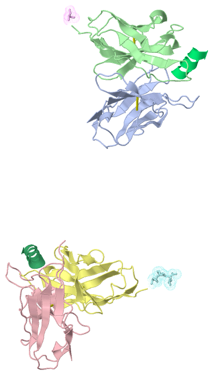
Chain A from PDB  Type:PROTEIN  Length:115
                                                                                                                                                    
               SCOP domains d2ap2a_ A: Immunoglobulin light chain kappa variable domain, VL-kappa                                               SCOP domains
               CATH domains 2ap2A00 A:-2-112 Immunoglobulins                                                                                    CATH domains
               Pfam domains ------------------------------------------------------------------------------------------------------------------- Pfam domains
         Sec.struct. author ......eeee..eee......eeeeeee.............eeeeee......eeeee............eeeeee..eeeeee....hhh..eeeeee............eee. Sec.struct. author
                 SAPs(SNPs) ------------------------------------------------------------------------------------------------------------------- SAPs(SNPs)
                    PROSITE ------------------------------------------------------------------------------------------------------------------- PROSITE
                 Transcript ------------------------------------------------------------------------------------------------------------------- Transcript
                2ap2 A   -2 FVRDIVMTQSPSSLTVTAGEKVTMSCKSSQSLLNSGNQKNYLTWYQQKPGQPPKLLIYWASTRESGVPDRFTGSGSGTDFTLTISSVQAEDLAVYYCQNDYSYPLTFGAGTKLEP  112
                                     7        17        27        37        47        57        67        77        87        97       107     

Chain B from PDB  Type:PROTEIN  Length:122
                                                                                                                                                           
               SCOP domains d2ap2b_ B: Immunoglobulin heavy chain variable domain, VH                                                                  SCOP domains
               CATH domains 2ap2B00 B:1-122 Immunoglobulins                                                                                            CATH domains
               Pfam domains -------------------------------------------------------------------------------------------------------------------------- Pfam domains
         Sec.struct. author ..eeee...........eeeeeeee...hhheeeeeeee.....eeeeeeee.....eee.......eeeeeehhh.eeeeee....hhh.eeeeeeeee..............eee..... Sec.struct. author
                 SAPs(SNPs) -------------------------------------------------------------------------------------------------------------------------- SAPs(SNPs)
                    PROSITE -------------------------------------------------------------------------------------------------------------------------- PROSITE
                 Transcript -------------------------------------------------------------------------------------------------------------------------- Transcript
                2ap2 B    1 EVQLQQSGAELVRPGASVKLSCTASGFNIKDDFMHWVKQRPEQGLEWIGRIDPANDNTKYAPKFQDKATIIADTSSNTAYLQLSSLTSEDTAVYYCARREVYSYYSPLDVWGAGTTVTVPSG  122
                                    10        20        30        40        50        60        70        80        90       100       110       120  

Chain C from PDB  Type:PROTEIN  Length:115
                                                                                                                                                    
               SCOP domains d2ap2c_ C: Immunoglobulin light chain kappa variable domain, VL-kappa                                               SCOP domains
               CATH domains 2ap2C00 C:-2-112 Immunoglobulins                                                                                    CATH domains
               Pfam domains ------------------------------------------------------------------------------------------------------------------- Pfam domains
         Sec.struct. author ......eeee..eee......eeeeeee.............eeeeee......eeee.....................eeeeee....hhh..eeeeee............eee. Sec.struct. author
                 SAPs(SNPs) ------------------------------------------------------------------------------------------------------------------- SAPs(SNPs)
                    PROSITE ------------------------------------------------------------------------------------------------------------------- PROSITE
                 Transcript ------------------------------------------------------------------------------------------------------------------- Transcript
                2ap2 C   -2 FVRDIVMTQSPSSLTVTAGEKVTMSCKSSQSLLNSGNQKNYLTWYQQKPGQPPKLLIYWASTRESGVPDRFTGSGSGTDFTLTISSVQAEDLAVYYCQNDYSYPLTFGAGTKLEP  112
                                     7        17        27        37        47        57        67        77        87        97       107     

Chain D from PDB  Type:PROTEIN  Length:122
                                                                                                                                                           
               SCOP domains d2ap2d_ D: Immunoglobulin heavy chain variable domain, VH                                                                  SCOP domains
               CATH domains 2ap2D00 D:1-122 Immunoglobulins                                                                                            CATH domains
               Pfam domains -------------------------------------------------------------------------------------------------------------------------- Pfam domains
         Sec.struct. author ..eeee...eee.....eeeeeeee...hhheeeeeeeee....eeeeeeee....eeee.hhh.............eeeeee........eeeeeeeee..............eeeee... Sec.struct. author
                 SAPs(SNPs) -------------------------------------------------------------------------------------------------------------------------- SAPs(SNPs)
                    PROSITE -------------------------------------------------------------------------------------------------------------------------- PROSITE
                 Transcript -------------------------------------------------------------------------------------------------------------------------- Transcript
                2ap2 D    1 EVQLQQSGAELVRPGASVKLSCTASGFNIKDDFMHWVKQRPEQGLEWIGRIDPANDNTKYAPKFQDKATIIADTSSNTAYLQLSSLTSEDTAVYYCARREVYSYYSPLDVWGAGTTVTVPSG  122
                                    10        20        30        40        50        60        70        80        90       100       110       120  

Chain E from PDB  Type:PROTEIN  Length:2
                                   
               SCOP domains -- SCOP domains
               CATH domains -- CATH domains
               Pfam domains -- Pfam domains
         Sec.struct. author .. Sec.struct. author
                 SAPs(SNPs) -- SAPs(SNPs)
                    PROSITE -- PROSITE
                 Transcript -- Transcript
                2ap2 E  123 SE  124

Chain F from PDB  Type:PROTEIN  Length:4
                                     
               SCOP domains ---- SCOP domains
               CATH domains ---- CATH domains
               Pfam domains ---- Pfam domains
         Sec.struct. author .... Sec.struct. author
                 SAPs(SNPs) ---- SAPs(SNPs)
                    PROSITE ---- PROSITE
                 Transcript ---- Transcript
                2ap2 F  123 SEQK  126

Chain P from PDB  Type:PROTEIN  Length:12
 aligned with MDR1_CRIGR | P21448 from UniProtKB/Swiss-Prot  Length:1276

    Alignment length:12
                                  1219  
          MDR1_CRIGR   1210 VVQEALDKAREG 1221
               SCOP domains ------------ SCOP domains
               CATH domains ------------ CATH domains
               Pfam domains ------------ Pfam domains
         Sec.struct. author hhhhhhhhhhh. Sec.struct. author
                 SAPs(SNPs) ------------ SAPs(SNPs)
                    PROSITE ------------ PROSITE
                 Transcript ------------ Transcript
                2ap2 P    1 VVQEALDKAREG   12
                                    10  

Chain Q from PDB  Type:PROTEIN  Length:14
 aligned with MDR1_CRIGR | P21448 from UniProtKB/Swiss-Prot  Length:1276

    Alignment length:14
                                  1219    
          MDR1_CRIGR   1210 VVQEALDKAREGRT 1223
               SCOP domains -------------- SCOP domains
               CATH domains -------------- CATH domains
               Pfam domains -------------- Pfam domains
         Sec.struct. author hhhhhhhhhhhhhh Sec.struct. author
                 SAPs(SNPs) -------------- SAPs(SNPs)
                    PROSITE -------------- PROSITE
                 Transcript -------------- Transcript
                2ap2 Q    1 VVQEALDKAREGRT   14
                                    10    

   Legend:   → Mismatch (orange background)
  - → Gap (green background, '-', border residues have a numbering label)
    → Modified Residue (blue background, lower-case, 'x' indicates undefined single-letter code, labelled with number + name)
  x → Chemical Group (purple background, 'x', labelled with number + name, e.g. ACE or NH2)
  extra numbering lines below/above indicate numbering irregularities and modified residue names etc., number ends below/above '|'

 Classification and Annotation

(-) SCOP Domains  (2, 4)

Asymmetric/Biological Unit

(-) CATH Domains  (1, 4)

Asymmetric/Biological Unit
(-)
Class: Mainly Beta (13760)

(-) Pfam Domains  (0, 0)

(no "Pfam Domain" information available for 2AP2)

(-) Gene Ontology  (13, 13)

Asymmetric/Biological Unit(hide GO term definitions)
Chain P,Q   (MDR1_CRIGR | P21448)
molecular function
    GO:0005524    ATP binding    Interacting selectively and non-covalently with ATP, adenosine 5'-triphosphate, a universally important coenzyme and enzyme regulator.
    GO:0016887    ATPase activity    Catalysis of the reaction: ATP + H2O = ADP + phosphate + 2 H+. May or may not be coupled to another reaction.
    GO:0042626    ATPase activity, coupled to transmembrane movement of substances    Catalysis of the reaction: ATP + H2O = ADP + phosphate, to directly drive the active transport of a substance across a membrane.
    GO:0016787    hydrolase activity    Catalysis of the hydrolysis of various bonds, e.g. C-O, C-N, C-C, phosphoric anhydride bonds, etc. Hydrolase is the systematic name for any enzyme of EC class 3.
    GO:0000166    nucleotide binding    Interacting selectively and non-covalently with a nucleotide, any compound consisting of a nucleoside that is esterified with (ortho)phosphate or an oligophosphate at any hydroxyl group on the ribose or deoxyribose.
    GO:0008559    xenobiotic-transporting ATPase activity    Catalysis of the reaction: ATP + H2O + xenobiotic(in) = ADP + phosphate + xenobiotic(out).
biological process
    GO:0006855    drug transmembrane transport    The process in which a drug is transported from one side of a membrane to the other by means of some agent such as a transporter or pore.
    GO:0055085    transmembrane transport    The process in which a solute is transported across a lipid bilayer, from one side of a membrane to the other
    GO:0006810    transport    The directed movement of substances (such as macromolecules, small molecules, ions) or cellular components (such as complexes and organelles) into, out of or within a cell, or between cells, or within a multicellular organism by means of some agent such as a transporter, pore or motor protein.
    GO:0042908    xenobiotic transport    The directed movement of a xenobiotic, a compound foreign to living organisms, into, out of or within a cell, or between cells, by means of some agent such as a transporter or pore.
cellular component
    GO:0016021    integral component of membrane    The component of a membrane consisting of the gene products and protein complexes having at least some part of their peptide sequence embedded in the hydrophobic region of the membrane.
    GO:0016020    membrane    A lipid bilayer along with all the proteins and protein complexes embedded in it an attached to it.
    GO:0005886    plasma membrane    The membrane surrounding a cell that separates the cell from its external environment. It consists of a phospholipid bilayer and associated proteins.

 Visualization

(-) Interactive Views

Asymmetric/Biological Unit
  Complete Structure
    Jena3D(integrated viewing of ligand, site, SAP, PROSITE, SCOP information)
    WebMol | AstexViewer[tm]@PDBe
(Java Applets, require no local installation except for Java; loading may be slow)
    STRAP
(Java WebStart application, automatic local installation, requires Java; full application with system access!)
    RasMol
(require local installation)
    Molscript (VRML)
(requires installation of a VRML viewer; select preferred view via VRML and generate a mono or stereo PDF format file)
 
  Ligands, Modified Residues, Ions
(no "Ligands, Modified Residues, Ions" information available for 2ap2)
 
  Sites
(no "Sites" information available for 2ap2)
 
  Cis Peptide Bonds
    Ser A:7 - Pro A:8   [ RasMol ]  
    Ser C:7 - Pro C:8   [ RasMol ]  
    Tyr A:100 - Pro A:101   [ RasMol ]  
    Tyr C:100 - Pro C:101   [ RasMol ]  
 

(-) Still Images

Jmol
  protein: cartoon or spacefill or dots and stick; nucleic acid: cartoon and stick; ligands: spacefill; active site: stick
Molscript
  protein, nucleic acid: cartoon; ligands: spacefill; active site: ball and stick

 Databases and Analysis Tools

(-) Databases

Access by PDB/NDB ID
  2ap2
    Family and Domain InformationProDom | SYSTERS
    General Structural InformationGlycoscienceDB | MMDB | NDB | OCA | PDB | PDBe | PDBj | PDBsum | PDBWiki | PQS | PROTEOPEDIA
    Orientation in MembranesOPM
    Protein SurfaceSURFACE
    Secondary StructureDSSP (structure derived) | HSSP (homology derived)
    Structural GenomicsGeneCensus
    Structural NeighboursCE | VAST
    Structure ClassificationCATH | Dali | SCOP
    Validation and Original DataBMRB Data View | BMRB Restraints Grid | EDS | PROCHECK | RECOORD | WHAT_CHECK
 
Access by UniProt ID/Accession number
  MDR1_CRIGR | P21448
    Comparative Protein Structure ModelsModBase
    Genomic InformationEnsembl
    Protein-protein InteractionDIP
    Sequence, Family and Domain InformationInterPro | Pfam | SMART | UniProtKB/SwissProt
 
Access by Enzyme Classificator   (EC Number)
  (no 'Enzyme Classificator' available)
    General Enzyme InformationBRENDA | EC-PDB | Enzyme | IntEnz
    PathwayKEGG | MetaCyc
 
Access by Disease Identifier   (MIM ID)
  (no 'MIM ID' available)
    Disease InformationOMIM
 
Access by GenAge ID
  (no 'GenAge ID' available)
    Age Related InformationGenAge

(-) Analysis Tools

Access by PDB/NDB ID
    Domain InformationXDom
    Interatomic Contacts of Structural UnitsCSU
    Ligand-protein ContactsLPC
    Protein CavitiescastP
    Sequence and Secondary StructurePDBCartoon
    Structure AlignmentSTRAP(Java WebStart application, automatic local installation, requires Java; full application with system access!)
    Structure and Sequence BrowserSTING
 
Access by UniProt ID/Accession number
  MDR1_CRIGR | P21448
    Protein Disorder PredictionDisEMBL | FoldIndex | GLOBPLOT (for more information see DisProt)

 Related Entries

(-) Entries Sharing at Least One Protein Chain (UniProt ID)

UniProtKB/Swiss-Prot
        MDR1_CRIGR | P214481mvu

(-) Related Entries Specified in the PDB File

(no "Related Entries Specified in the PDB File" available for 2AP2)