Show PDB file:   
         Plain Text   HTML   (compressed file size)
QuickSearch:   
by PDB,NDB,UniProt,PROSITE Code or Search Term(s)  
(-)NMR Structure - manually
(-)NMR Structure - model 1
(-)NMR Structure - all models
collapse expand < >
Image NMR Structure - manually
NMR Structure - manually  (Jmol Viewer)
Image NMR Structure - model 1
NMR Structure - model 1  (Jmol Viewer)
Image NMR Structure - all models
NMR Structure - all models  (Jmol Viewer)

(-) Description

Title :  SOLUTION STRUCTURE OF THE JOINED PH DOMAIN OF ALPHA1-SYNTROPHIN
 
Authors :  J. Yan, W. Wen, W. Xu, J. F. Long, M. E. Adams, S. C. Froehner, M. Zhang
Date :  21 Jul 05  (Deposition) - 24 Jan 06  (Release) - 24 Feb 09  (Revision)
Method :  SOLUTION NMR
Resolution :  NOT APPLICABLE
Chains :  NMR Structure  :  A  (20x)
Keywords :  Protein Binding (Keyword Search: [Gene Ontology, PubMed, Web (Google))
 
Reference :  J. Yan, W. Wen, W. Xu, J. F. Long, M. E. Adams, S. C. Froehner, M. Zhang
Structure Of The Split Ph Domain And Distinct Lipid-Binding Properties Of The Ph-Pdz Supramodule Of Alpha-Syntrophin
Embo J. V. 24 3985 2005
PubMed-ID: 16252003  |  Reference-DOI: 10.1038/SJ.EMBOJ.7600858
(for further references see the PDB file header)

(-) Compounds

Molecule 1 - ALPHA-1-SYNTROPHIN
    ChainsA
    EngineeredYES
    Expression SystemESCHERICHIA COLI
    Expression System PlasmidPET32A(A MODIFIED VERSION)
    Expression System Taxid562
    Expression System Vector TypePLASMID
    FragmentPH DOMAIN
    Organism CommonHOUSE MOUSE
    Organism ScientificMUS MUSCULUS
    Organism Taxid10090
    SynonymALPHA-SYNTROPHIN

 Structural Features

(-) Chains, Units

  
NMR Structure (20x)

Summary Information (see also Sequences/Alignments below)

(-) Ligands, Modified Residues, Ions  (0, 0)

(no "Ligand,Modified Residues,Ions" information available for 2ADZ)

(-) Sites  (0, 0)

(no "Site" information available for 2ADZ)

(-) SS Bonds  (0, 0)

(no "SS Bond" information available for 2ADZ)

(-) Cis Peptide Bonds  (0, 0)

(no "Cis Peptide Bond" information available for 2ADZ)

 Sequence-Structure Mapping

(-) SAPs(SNPs)/Variants  (0, 0)

(no "SAP(SNP)/Variant" information available for 2ADZ)

(-) PROSITE Motifs  (0, 0)

(no "PROSITE Motif" information available for 2ADZ)

(-) Exons   (0, 0)

(no "Exon" information available for 2ADZ)

(-) Sequences/Alignments

NMR Structure
   Reformat: Number of residues per line =  ('0' or empty: single-line sequence representation)
  Number of residues per labelling interval =   
  UniProt sequence: complete  aligned part    
   Show mapping: SCOP domains CATH domains Pfam domains Secondary structure (by author)
SAPs(SNPs) PROSITE motifs Exons
(details for a mapped element are shown in a popup box when the mouse pointer rests over it)
Chain A from PDB  Type:PROTEIN  Length:178
 aligned with SNTA1_MOUSE | Q61234 from UniProtKB/Swiss-Prot  Length:503

    Alignment length:263
                                    11        21        31        41        51        61        71        81        91       101       111       121       131       141       151       161       171       181       191       201       211       221       231       241       251       261   
          SNTA1_MOUSE     2 ASGRRAPRTGLLELRCGAGSGAGGERWQRVLLSLAEDALTVSPADGEPGPEPEPAQLNGAAEPGAAPPQLPEALLLQRRRVTVRKADAGGLGISIKGGRENKMPILISKIFKGLAADQTEALFVGDAILSVNGEDLSSATHDEAVQALKKTGKEVVLEVKYMKEVSPYFKNSAGGTSVGWDSPPASPLQRQPSSPGPQPRNLSEAKHVSLKMAYVSRRCTPTDPEPRYLEICAADGQDAVFLRAKDEASARSWAGAIQAQIGT 264
               SCOP domains d2adza1 A:1-43,A:117-178 Alpha-1-syntrophin--------------------------------------------------------------------------------------------------------------------------------------------------------------d2adza1 A:1-43,A:117-178 Alpha-1-syntrophin                    SCOP domains
               CATH domains ----------------------------------------------------------------------------------------------------------------------------------------------------------------------------------------------------------------------------------------------------------------------- CATH domains
               Pfam domains ----------------------------------------------------------------------------------------------------------------------------------------------------------------------------------------------------------------------------------------------------------------------- Pfam domains
         Sec.struct. author .......eeeeeee.............eeeeeeee..eeeee...............................hhhhh-------------------------------------------------------------------------------------hh........................................eee.....eeeee.........eeeeee....eeeeee..hhhhhhhhhhhhhhhhhh Sec.struct. author
                 SAPs(SNPs) ----------------------------------------------------------------------------------------------------------------------------------------------------------------------------------------------------------------------------------------------------------------------- SAPs(SNPs)
                    PROSITE ----------------------------------------------------------------------------------------------------------------------------------------------------------------------------------------------------------------------------------------------------------------------- PROSITE
                 Transcript ----------------------------------------------------------------------------------------------------------------------------------------------------------------------------------------------------------------------------------------------------------------------- Transcript
                 2adz A   1 ASGRRAPRTGLLELRCGAGSGAGGERWQRVLLSLAEDALTVSPADGEPGPEPEPAQLNGAAEPGAAPPQLPEALLLQR-------------------------------------------------------------------------------------EVSPYFKNSAGGTSVGWDSPPASPLQRQPSSPGPQPRNLSEAKHVSLKMAYVSRRCTPTDPEPRYLEICAADGQDAVFLRAKDEASARSWAGAIQAQIGT 178
                                    10        20        30        40        50        60        70       | -         -         -         -         -         -         -         -         -   |    85        95       105       115       125       135       145       155       165       175   
                                                                                                        78                                                                                    79                                                                                                   

   Legend:   → Mismatch (orange background)
  - → Gap (green background, '-', border residues have a numbering label)
    → Modified Residue (blue background, lower-case, 'x' indicates undefined single-letter code, labelled with number + name)
  x → Chemical Group (purple background, 'x', labelled with number + name, e.g. ACE or NH2)
  extra numbering lines below/above indicate numbering irregularities and modified residue names etc., number ends below/above '|'

 Classification and Annotation

(-) SCOP Domains  (1, 1)

NMR Structure

(-) CATH Domains  (0, 0)

(no "CATH Domain" information available for 2ADZ)

(-) Pfam Domains  (0, 0)

(no "Pfam Domain" information available for 2ADZ)

(-) Gene Ontology  (24, 24)

NMR Structure(hide GO term definitions)
Chain A   (SNTA1_MOUSE | Q61234)
molecular function
    GO:0051117    ATPase binding    Interacting selectively and non-covalently with an ATPase, any enzyme that catalyzes the hydrolysis of ATP.
    GO:0003779    actin binding    Interacting selectively and non-covalently with monomeric or multimeric forms of actin, including actin filaments.
    GO:0005516    calmodulin binding    Interacting selectively and non-covalently with calmodulin, a calcium-binding protein with many roles, both in the calcium-bound and calcium-free states.
    GO:0044325    ion channel binding    Interacting selectively and non-covalently with one or more specific sites on an ion channel, a protein complex that spans a membrane and forms a water-filled channel across the phospholipid bilayer allowing selective ion transport down its electrochemical gradient.
    GO:0050998    nitric-oxide synthase binding    Interacting selectively and non-covalently with the enzyme nitric-oxide synthase.
    GO:0005515    protein binding    Interacting selectively and non-covalently with any protein or protein complex (a complex of two or more proteins that may include other nonprotein molecules).
    GO:0017080    sodium channel regulator activity    Modulates the activity of a sodium channel.
    GO:0005198    structural molecule activity    The action of a molecule that contributes to the structural integrity of a complex or its assembly within or outside a cell.
biological process
    GO:1902083    negative regulation of peptidyl-cysteine S-nitrosylation    Any process that stops, prevents or reduces the frequency, rate or extent of peptidyl-cysteine S-nitrosylation.
    GO:0007528    neuromuscular junction development    A process that is carried out at the cellular level which results in the assembly, arrangement of constituent parts, or disassembly of a neuromuscular junction.
    GO:0002027    regulation of heart rate    Any process that modulates the frequency or rate of heart contraction.
    GO:1902305    regulation of sodium ion transmembrane transport    Any process that modulates the frequency, rate or extent of sodium ion transmembrane transport.
    GO:0003117    regulation of vasoconstriction by circulating norepinephrine    Any process that modulates the frequency, rate or extent of reductions in the diameter of blood vessels as a result of secretion of norepinephrine into the bloodstream.
    GO:0060307    regulation of ventricular cardiac muscle cell membrane repolarization    Any process that modulates the establishment or extent of a membrane potential in the polarizing direction towards the resting potential in a ventricular cardiomyocyte.
    GO:0086005    ventricular cardiac muscle cell action potential    An action potential that occurs in a ventricular cardiac muscle cell.
cellular component
    GO:0030054    cell junction    A cellular component that forms a specialized region of connection between two or more cells or between a cell and the extracellular matrix. At a cell junction, anchoring proteins extend through the plasma membrane to link cytoskeletal proteins in one cell to cytoskeletal proteins in neighboring cells or to proteins in the extracellular matrix.
    GO:0005737    cytoplasm    All of the contents of a cell excluding the plasma membrane and nucleus, but including other subcellular structures.
    GO:0005856    cytoskeleton    Any of the various filamentous elements that form the internal framework of cells, and typically remain after treatment of the cells with mild detergent to remove membrane constituents and soluble components of the cytoplasm. The term embraces intermediate filaments, microfilaments, microtubules, the microtrabecular lattice, and other structures characterized by a polymeric filamentous nature and long-range order within the cell. The various elements of the cytoskeleton not only serve in the maintenance of cellular shape but also have roles in other cellular functions, including cellular movement, cell division, endocytosis, and movement of organelles.
    GO:0005622    intracellular    The living contents of a cell; the matter contained within (but not including) the plasma membrane, usually taken to exclude large vacuoles and masses of secretory or ingested material. In eukaryotes it includes the nucleus and cytoplasm.
    GO:0016020    membrane    A lipid bilayer along with all the proteins and protein complexes embedded in it an attached to it.
    GO:0005886    plasma membrane    The membrane surrounding a cell that separates the cell from its external environment. It consists of a phospholipid bilayer and associated proteins.
    GO:0045211    postsynaptic membrane    A specialized area of membrane facing the presynaptic membrane on the tip of the nerve ending and separated from it by a minute cleft (the synaptic cleft). Neurotransmitters cross the synaptic cleft and transmit the signal to the postsynaptic membrane.
    GO:0043234    protein complex    A stable macromolecular complex composed (only) of two or more polypeptide subunits along with any covalently attached molecules (such as lipid anchors or oligosaccharide) or non-protein prosthetic groups (such as nucleotides or metal ions). Prosthetic group in this context refers to a tightly bound cofactor. The component polypeptide subunits may be identical.
    GO:0042383    sarcolemma    The outer membrane of a muscle cell, consisting of the plasma membrane, a covering basement membrane (about 100 nm thick and sometimes common to more than one fiber), and the associated loose network of collagen fibers.
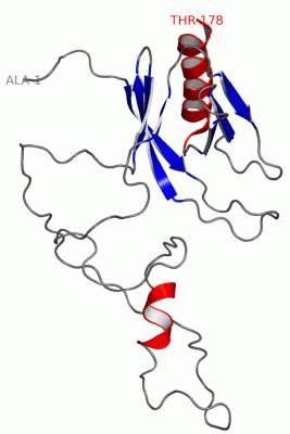
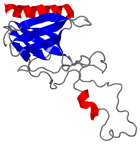
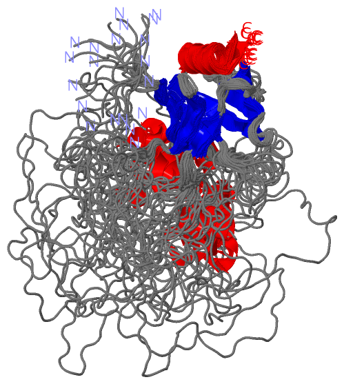
 Visualization

(-) Interactive Views

NMR Structure
  Complete Structure
    Jena3D(integrated viewing of ligand, site, SAP, PROSITE, SCOP information)
    WebMol | AstexViewer[tm]@PDBe
(Java Applets, require no local installation except for Java; loading may be slow)
    STRAP
(Java WebStart application, automatic local installation, requires Java; full application with system access!)
    RasMol
(require local installation)
    Molscript (VRML)
(requires installation of a VRML viewer; select preferred view via VRML and generate a mono or stereo PDF format file)
 
  Ligands, Modified Residues, Ions
(no "Ligands, Modified Residues, Ions" information available for 2adz)
 
  Sites
(no "Sites" information available for 2adz)
 
  Cis Peptide Bonds
(no "Cis Peptide Bonds" information available for 2adz)
 

(-) Still Images

Jmol
  protein: cartoon or spacefill or dots and stick; nucleic acid: cartoon and stick; ligands: spacefill; active site: stick
Molscript
  protein, nucleic acid: cartoon; ligands: spacefill; active site: ball and stick

 Databases and Analysis Tools

(-) Databases

Access by PDB/NDB ID
  2adz
    Family and Domain InformationProDom | SYSTERS
    General Structural InformationGlycoscienceDB | MMDB | NDB | OCA | PDB | PDBe | PDBj | PDBsum | PDBWiki | PQS | PROTEOPEDIA
    Orientation in MembranesOPM
    Protein SurfaceSURFACE
    Secondary StructureDSSP (structure derived) | HSSP (homology derived)
    Structural GenomicsGeneCensus
    Structural NeighboursCE | VAST
    Structure ClassificationCATH | Dali | SCOP
    Validation and Original DataBMRB Data View | BMRB Restraints Grid | EDS | PROCHECK | RECOORD | WHAT_CHECK
 
Access by UniProt ID/Accession number
  SNTA1_MOUSE | Q61234
    Comparative Protein Structure ModelsModBase
    Genomic InformationEnsembl
    Protein-protein InteractionDIP
    Sequence, Family and Domain InformationInterPro | Pfam | SMART | UniProtKB/SwissProt
 
Access by Enzyme Classificator   (EC Number)
  (no 'Enzyme Classificator' available)
    General Enzyme InformationBRENDA | EC-PDB | Enzyme | IntEnz
    PathwayKEGG | MetaCyc
 
Access by Disease Identifier   (MIM ID)
  (no 'MIM ID' available)
    Disease InformationOMIM
 
Access by GenAge ID
  (no 'GenAge ID' available)
    Age Related InformationGenAge

(-) Analysis Tools

Access by PDB/NDB ID
    Domain InformationXDom
    Interatomic Contacts of Structural UnitsCSU
    Ligand-protein ContactsLPC
    Protein CavitiescastP
    Sequence and Secondary StructurePDBCartoon
    Structure AlignmentSTRAP(Java WebStart application, automatic local installation, requires Java; full application with system access!)
    Structure and Sequence BrowserSTING
 
Access by UniProt ID/Accession number
  SNTA1_MOUSE | Q61234
    Protein Disorder PredictionDisEMBL | FoldIndex | GLOBPLOT (for more information see DisProt)

 Related Entries

(-) Entries Sharing at Least One Protein Chain (UniProt ID)

UniProtKB/Swiss-Prot
        SNTA1_MOUSE | Q612341qav 1z86 1z87 2pdz 4hop

(-) Related Entries Specified in the PDB File

1z87 THE ISOLATED PH DOMAIN OF THE PROTEIN