|
|
|
|
 Description
|
|
Compounds
|
||||||||||||||||||||
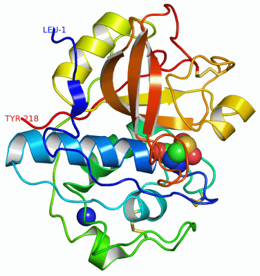
Chains, Units
Summary Information (see also Sequences/Alignments below) |
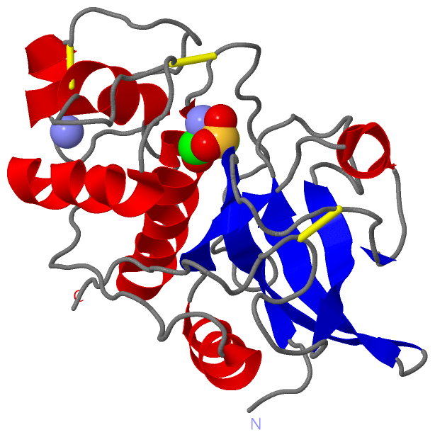
Ligands, Modified Residues, Ions (2, 2)| Asymmetric/Biological Unit (2, 2) |
Sites (3, 3)
Asymmetric Unit (3, 3)
|
SS Bonds (3, 3)
Asymmetric/Biological Unit
|
||||||||||||||||
Cis Peptide Bonds (1, 1)
Asymmetric/Biological Unit
|
||||||||
SAPs(SNPs)/Variants (0, 0)| (no "SAP(SNP)/Variant" information available for 2ACT) |
PROSITE Motifs (3, 3)
Asymmetric/Biological Unit (3, 3)
|
||||||||||||||||||||||||||||||||||||||||
Exons (0, 0)| (no "Exon" information available for 2ACT) |
Sequences/Alignments
Asymmetric/Biological UnitChain A from PDB Type:PROTEIN Length:218 aligned with ACTN_ACTCH | P00785 from UniProtKB/Swiss-Prot Length:380 Alignment length:218 136 146 156 166 176 186 196 206 216 226 236 246 256 266 276 286 296 306 316 326 336 ACTN_ACTCH 127 LPSYVDWRSAGAVVDIKSQGECGGCWAFSAIATVEGINKIVTGVLISLSEQELIDCGRTQNTRGCNGGYITDGFQFIINNGGINTEENYPYTAQDGECNVDLQNEKYVTIDTYENVPYNNEWALQTAVTYQPVSVALDAAGDAFKQYSSGIFTGPCGTAVDHAVTIVGYGTEGGIDYWIVKNSWDTTWGEEGYMRILRNVGGAGTCGIATMPSYPVKY 344 SCOP domains d2acta_ A: Actinidin SCOP domains CATH domains 2actA00 A:1-218 Cysteine proteinases CATH domains Pfam domains -------------------------------------------------------------------------------------------------------------------------------------------------------------------------------------------------------------------------- Pfam domains SAPs(SNPs) -------------------------------------------------------------------------------------------------------------------------------------------------------------------------------------------------------------------------- SAPs(SNPs) PROSITE ------------------THIOL_PROTEA---------------------------------------------------------------------------------------------------------------------------------THIOL_PROTE------THIOL_PROTEASE_ASN ---------------------- PROSITE Transcript -------------------------------------------------------------------------------------------------------------------------------------------------------------------------------------------------------------------------- Transcript 2act A 1 LPSYVDWRSAGAVVDIKSQGECGGcWAFSAIATVEGINKITSGSLISLSEQELIDCGRTQNTRGCDGGYITDGFQFIINDGGINTEENYPYTAQDGDCDVALQDQKYVTIDTYENVPYNNEWALQTAVTYQPVSVALDAAGDAFKQYASGIFTGPCGTAVDHAIVIVGYGTEGGVDYWIVKNSWDTTWGEEGYMRILRNVGGAGTCGIATMPSYPVKY 218 10 20 | 30 40 50 60 70 80 90 100 110 120 130 140 150 160 170 180 190 200 210 25-CSD
|
||||||||||||||||||||
SCOP Domains (1, 1)
Asymmetric/Biological Unit
 |
CATH Domains (1, 1)
Asymmetric/Biological Unit
|
Pfam Domains (0, 0)| (no "Pfam Domain" information available for 2ACT) |
Gene Ontology (4, 4)|
Asymmetric/Biological Unit(hide GO term definitions) Chain A (ACTN_ACTCH | P00785)
|
||||||||||||||||||||||||||||||||||||
Interactive Views
|
||||||||||||||||||||||||||||||||||||||||||||||||||||||||||||||||||||||||||||||||||||||||||||||||||||||||||||||||||||||||||||||||||||||||||||
Still Images
|
||||||||||||||||
Databases
|
||||||||||||||||||||||||||||||||||||||||||||||||||||||||||||||||||||||||||||||||||||||||||||||||||||||||||||||||||||||||||||||||||||||||||||||||||||||||||||||||
Analysis Tools
|
|||||||||||||||||||||||||||||||||||||||||||||||||||||||||||||
Entries Sharing at Least One Protein Chain (UniProt ID)
Related Entries Specified in the PDB File
|
|