Show PDB file:   
         Plain Text   HTML   (compressed file size)
QuickSearch:   
by PDB,NDB,UniProt,PROSITE Code or Search Term(s)  
(-)Asym./Biol. Unit
(-)Asym./Biol. Unit - sites
collapse expand < >
Image Asym./Biol. Unit
Asym./Biol. Unit  (Jmol Viewer)
Image Asym./Biol. Unit - sites
Asym./Biol. Unit - sites  (Jmol Viewer)

(-) Description

Title :  CRYSTAL STRUCTURE OF PROTEIN-GLUTAMINASE
 
Authors :  R. Hashizume
Date :  13 Mar 08  (Deposition) - 17 Mar 09  (Release) - 29 Aug 12  (Revision)
Method :  X-RAY DIFFRACTION
Resolution :  1.15
Chains :  Asym./Biol. Unit :  X
Keywords :  Deamidation Glutaminase, Hydrolase (Keyword Search: [Gene Ontology, PubMed, Web (Google))
 
Reference :  R. Hashizume, Y. Maki, K. Mizutani, N. Takahashi, H. Matsubara, A. Sugita, K. Sato, S. Yamaguchi, B. Mikami
Crystal Structures Of Protein Glutaminase And Its Pro Forms Converted Into Enzyme-Substrate Complex
J. Biol. Chem. V. 286 38691 2011
PubMed-ID: 21926168  |  Reference-DOI: 10.1074/JBC.M111.255133

(-) Compounds

Molecule 1 - PROTEIN-GLUTAMINASE
    ChainsX
    EC Number3.5.1.-
    EngineeredYES
    Expression SystemESCHERICHIA COLI
    Expression System PlasmidPN7-9
    Expression System Taxid562
    Expression System Vector TypePLASMID
    Organism ScientificCHRYSEOBACTERIUM PROTEOLYTICUM
    Organism Taxid118127
    Strain9670

 Structural Features

(-) Chains, Units

  1
Asymmetric/Biological Unit X

Summary Information (see also Sequences/Alignments below)
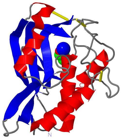
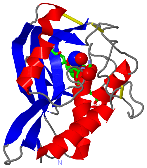
(-) Ligands, Modified Residues, Ions  (2, 2)

Asymmetric/Biological Unit (2, 2)
No.NameCountTypeFull Name
1GOL1Ligand/IonGLYCEROL
2NA1Ligand/IonSODIUM ION

(-) Sites  (4, 4)

Asymmetric Unit (4, 4)
No.NameEvidenceResiduesDescription
1AC1unknownGLY X:41 , CYS X:42 , HOH X:565 , HOH X:619 , HOH X:789BINDING SITE FOR RESIDUE NA X 501
2AC2unknownTYR X:43 , TYR X:82 , PHE X:185 , HOH X:642 , HOH X:703BINDING SITE FOR RESIDUE GOL X 201
3AC3SOFTWAREGLY X:41 , CYS X:42 , HOH X:565 , HOH X:619 , HOH X:789BINDING SITE FOR RESIDUE NA X 501
4AC4SOFTWARETYR X:43 , TYR X:82 , PHE X:185 , HOH X:642 , HOH X:703BINDING SITE FOR RESIDUE GOL X 201

(-) SS Bonds  (4, 4)

Asymmetric/Biological Unit
No.Residues
1X:23 -X:32
2X:76 -X:172
3X:77 -X:126
4X:161 -X:183

(-) Cis Peptide Bonds  (3, 3)

Asymmetric/Biological Unit
No.Residues
1Ser X:30 -Pro X:31
2Ser X:173 -Pro X:174
3Ser X:175 -Pro X:176

 Sequence-Structure Mapping

(-) SAPs(SNPs)/Variants  (0, 0)

(no "SAP(SNP)/Variant" information available for 2ZK9)

(-) PROSITE Motifs  (0, 0)

(no "PROSITE Motif" information available for 2ZK9)

(-) Exons   (0, 0)

(no "Exon" information available for 2ZK9)

(-) Sequences/Alignments

Asymmetric/Biological Unit
   Reformat: Number of residues per line =  ('0' or empty: single-line sequence representation)
  Number of residues per labelling interval =   
  UniProt sequence: complete  aligned part    
   Show mapping: SCOP domains CATH domains Pfam domains Secondary structure (by author)
SAPs(SNPs) PROSITE motifs Exons
(details for a mapped element are shown in a popup box when the mouse pointer rests over it)
Chain X from PDB  Type:PROTEIN  Length:185
 aligned with Q9AQQ8_9FLAO | Q9AQQ8 from UniProtKB/TrEMBL  Length:320

    Alignment length:185
                                   145       155       165       175       185       195       205       215       225       235       245       255       265       275       285       295       305       315     
         Q9AQQ8_9FLAO   136 LASVIPDVATLNSLFNQIKNQSCGTSTASSPCITFRYPVDGCYARAHKMRQILMNNGYDCEKQFVYGNLKASTGTCCVAWSYHVAILVSYKNASGVTEKRIIDPSLFSSGPVTDTAWRNACVNTSCGSASVSSYANTAGNVYYRSPSNSYLYDNNLINTNCVLTKFSLLSGCSPSPAPDVSSCGF 320
               SCOP domains ----------------------------------------------------------------------------------------------------------------------------------------------------------------------------------------- SCOP domains
               CATH domains ----------------------------------------------------------------------------------------------------------------------------------------------------------------------------------------- CATH domains
               Pfam domains ----------------------------------------------------------------------------------------------------------------------------------------------------------------------------------------- Pfam domains
         Sec.struct. author ...ee.hhhhhhhhhhhhhh....................hhhhhhhhhhhhhhhh....eeeeeeeeeeee....eeeeeeeeeeeeeee.....eeeeee..........hhhhhhhhhhhhhhh..eeeeeeeee..eeee.....eee...hhhhhhhhhhh................... Sec.struct. author
                 SAPs(SNPs) ----------------------------------------------------------------------------------------------------------------------------------------------------------------------------------------- SAPs(SNPs)
                    PROSITE ----------------------------------------------------------------------------------------------------------------------------------------------------------------------------------------- PROSITE
                 Transcript ----------------------------------------------------------------------------------------------------------------------------------------------------------------------------------------- Transcript
                 2zk9 X   1 LASVIPDVATLNSLFNQIKNESCGTSTASSPCITFRYPVDGCYARAHKMRQILMNNGYDCEKQFVYGNLKASTGTCCVAWSYHVAILVSYKNASGVTEKRIIDPSLFSSGPVTDTAWRNACVNTSCGSASVSSYANTAGNVYYRSPSNSYLYDNNLINTNCVLTKFSLLSGCSPSPAPDVSSCGF 185
                                    10        20        30        40        50        60        70        80        90       100       110       120       130       140       150       160       170       180     

   Legend:   → Mismatch (orange background)
  - → Gap (green background, '-', border residues have a numbering label)
    → Modified Residue (blue background, lower-case, 'x' indicates undefined single-letter code, labelled with number + name)
  x → Chemical Group (purple background, 'x', labelled with number + name, e.g. ACE or NH2)
  extra numbering lines below/above indicate numbering irregularities and modified residue names etc., number ends below/above '|'

 Classification and Annotation

(-) SCOP Domains  (0, 0)

(no "SCOP Domain" information available for 2ZK9)

(-) CATH Domains  (0, 0)

(no "CATH Domain" information available for 2ZK9)

(-) Pfam Domains  (0, 0)

(no "Pfam Domain" information available for 2ZK9)

(-) Gene Ontology  (0, 0)

Asymmetric/Biological Unit(hide GO term definitions)
    (no "Gene Ontology" information available for 2ZK9)

 Visualization

(-) Interactive Views

Asymmetric/Biological Unit
  Complete Structure
    Jena3D(integrated viewing of ligand, site, SAP, PROSITE, SCOP information)
    WebMol | AstexViewer[tm]@PDBe
(Java Applets, require no local installation except for Java; loading may be slow)
    STRAP
(Java WebStart application, automatic local installation, requires Java; full application with system access!)
    RasMol
(require local installation)
    Molscript (VRML)
(requires installation of a VRML viewer; select preferred view via VRML and generate a mono or stereo PDF format file)
 
  Ligands, Modified Residues, Ions
    GOL  [ RasMol | Jena3D ]  +environment [ RasMol | Jena3D ]
    NA  [ RasMol | Jena3D ]  +environment [ RasMol | Jena3D ]
 
  Sites
    AC1  [ RasMol ]  +environment [ RasMol ]
    AC2  [ RasMol ]  +environment [ RasMol ]
    AC3  [ RasMol ]  +environment [ RasMol ]
    AC4  [ RasMol ]  +environment [ RasMol ]
 
  Cis Peptide Bonds
    Ser X:173 - Pro X:174   [ RasMol ]  
    Ser X:175 - Pro X:176   [ RasMol ]  
    Ser X:30 - Pro X:31   [ RasMol ]  
 

(-) Still Images

Jmol
  protein: cartoon or spacefill or dots and stick; nucleic acid: cartoon and stick; ligands: spacefill; active site: stick
Molscript
  protein, nucleic acid: cartoon; ligands: spacefill; active site: ball and stick

 Databases and Analysis Tools

(-) Databases

Access by PDB/NDB ID
  2zk9
    Family and Domain InformationProDom | SYSTERS
    General Structural InformationGlycoscienceDB | MMDB | NDB | OCA | PDB | PDBe | PDBj | PDBsum | PDBWiki | PQS | PROTEOPEDIA
    Orientation in MembranesOPM
    Protein SurfaceSURFACE
    Secondary StructureDSSP (structure derived) | HSSP (homology derived)
    Structural GenomicsGeneCensus
    Structural NeighboursCE | VAST
    Structure ClassificationCATH | Dali | SCOP
    Validation and Original DataBMRB Data View | BMRB Restraints Grid | EDS | PROCHECK | RECOORD | WHAT_CHECK
 
Access by UniProt ID/Accession number
  Q9AQQ8_9FLAO | Q9AQQ8
    Comparative Protein Structure ModelsModBase
    Genomic InformationEnsembl
    Protein-protein InteractionDIP
    Sequence, Family and Domain InformationInterPro | Pfam | SMART | UniProtKB/TrEMBL
 
Access by Enzyme Classificator   (EC Number)
  3.5.1.-
    General Enzyme InformationBRENDA | EC-PDB | Enzyme | IntEnz
    PathwayKEGG | MetaCyc
 
Access by Disease Identifier   (MIM ID)
  (no 'MIM ID' available)
    Disease InformationOMIM
 
Access by GenAge ID
  (no 'GenAge ID' available)
    Age Related InformationGenAge

(-) Analysis Tools

Access by PDB/NDB ID
    Domain InformationXDom
    Interatomic Contacts of Structural UnitsCSU
    Ligand-protein ContactsLPC
    Protein CavitiescastP
    Sequence and Secondary StructurePDBCartoon
    Structure AlignmentSTRAP(Java WebStart application, automatic local installation, requires Java; full application with system access!)
    Structure and Sequence BrowserSTING
 
Access by UniProt ID/Accession number
  Q9AQQ8_9FLAO | Q9AQQ8
    Protein Disorder PredictionDisEMBL | FoldIndex | GLOBPLOT (for more information see DisProt)

 Related Entries

(-) Entries Sharing at Least One Protein Chain (UniProt ID)

UniProtKB/TrEMBL
        Q9AQQ8_9FLAO | Q9AQQ82ksv 3a54 3a55 3a56

(-) Related Entries Specified in the PDB File

3a54 THE A47Q MUTANT OF THIS ENTRY
3a55 THE A47Q MUTANT OF THIS ENTRY
3a56 WILD PROTEIN OF THIS ENTRY