Show PDB file:   
         Plain Text   HTML   (compressed file size)
QuickSearch:   
by PDB,NDB,UniProt,PROSITE Code or Search Term(s)  
(-)Asymmetric Unit
(-)Asym. Unit - sites
(-)Biological Unit 1
(-)Biol. Unit 1 - sites
(-)Biological Unit 2
collapse expand < >
Image Asymmetric Unit
Asymmetric Unit  (Jmol Viewer)
Image Asym. Unit - sites
Asym. Unit - sites  (Jmol Viewer)
Image Biological Unit 1
Biological Unit 1  (Jmol Viewer)
Image Biol. Unit 1 - sites
Biol. Unit 1 - sites  (Jmol Viewer)
Image Biological Unit 2
Biological Unit 2  (Jmol Viewer)

(-) Description

Title :  ASN-HYDROXYLATION STABILISES THE ANKYRIN REPEAT DOMAIN FOLD
 
Authors :  M. A. Mcdonough, C. J. Schofield
Date :  21 Jan 08  (Deposition) - 05 Feb 08  (Release) - 27 Oct 09  (Revision)
Method :  X-RAY DIFFRACTION
Resolution :  1.90
Chains :  Asym. Unit :  A
Biol. Unit 1:  A  (1x)
Biol. Unit 2:  A  (2x)
Keywords :  Ankyrin Repeat, Hydroxylated, De Novo Protein (Keyword Search: [Gene Ontology, PubMed, Web (Google))
 
Reference :  L. Kelly, M. A. Mcdonough, M. L. Coleman, P. J. Ratcliffe, C. J. Schofield
Asparagine Beta-Hydroxylation Stabilizes The Ankyrin Repeat Domain Fold
Mol Biosyst V. 5 52 2009
PubMed-ID: 19081931  |  Reference-DOI: 10.1039/B815271C
(for further references see the PDB file header)

(-) Compounds

Molecule 1 - 3 REPEAT SYNTHETIC ANKYRIN
    ChainsA
    EngineeredYES
    Other DetailsSYNTHETICALLY DESIGNED CONSTRUCT
    SyntheticYES

 Structural Features

(-) Chains, Units

  1
Asymmetric Unit A
Biological Unit 1 (1x)A
Biological Unit 2 (2x)A

Summary Information (see also Sequences/Alignments below)

(-) Ligands, Modified Residues, Ions  (3, 8)

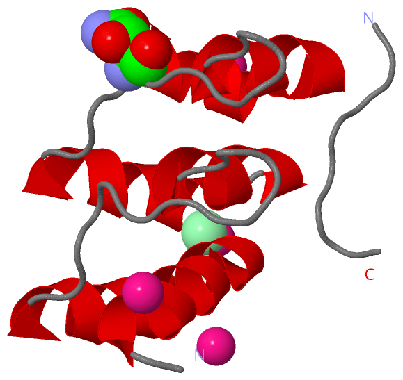
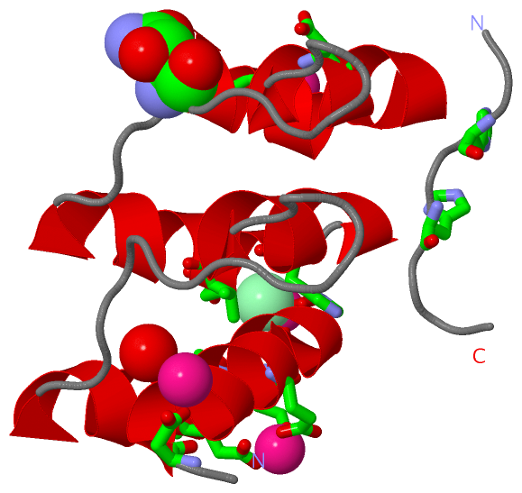
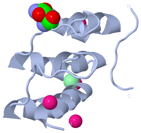
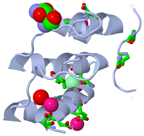
Asymmetric Unit (3, 8)
No.NameCountTypeFull Name
1AHB1Mod. Amino AcidBETA-HYDROXYASPARAGINE
2CD5Ligand/IonCADMIUM ION
3CL2Ligand/IonCHLORIDE ION
Biological Unit 1 (1, 1)
No.NameCountTypeFull Name
1AHB1Mod. Amino AcidBETA-HYDROXYASPARAGINE
2CD-1Ligand/IonCADMIUM ION
3CL-1Ligand/IonCHLORIDE ION
Biological Unit 2 (1, 2)
No.NameCountTypeFull Name
1AHB2Mod. Amino AcidBETA-HYDROXYASPARAGINE
2CD-1Ligand/IonCADMIUM ION
3CL-1Ligand/IonCHLORIDE ION

(-) Sites  (7, 7)

Asymmetric Unit (7, 7)
No.NameEvidenceResiduesDescription
1AC1SOFTWAREGLU A:30 , GLN A:36 , GLU A:39 , ASP A:95BINDING SITE FOR RESIDUE CD A 410
2AC2SOFTWAREHIS A:7 , HIS A:9 , GLU A:107 , HOH A:1018 , HOH A:1019BINDING SITE FOR RESIDUE CD A 411
3AC3SOFTWARECL A:415 , HOH A:1020 , HOH A:1050 , HOH A:1065 , HOH A:1066BINDING SITE FOR RESIDUE CD A 412
4AC4SOFTWAREASP A:37 , ASP A:38 , CL A:416 , HOH A:1046BINDING SITE FOR RESIDUE CD A 413
5AC5SOFTWAREASP A:23 , HOH A:1039 , HOH A:1045BINDING SITE FOR RESIDUE CD A 414
6AC6SOFTWAREHIS A:69 , GLU A:71 , VAL A:72 , CD A:412 , HOH A:1065 , HOH A:1066BINDING SITE FOR RESIDUE CL A 415
7AC7SOFTWAREGLN A:36 , ASP A:38 , GLU A:39 , CD A:413 , HOH A:1046BINDING SITE FOR RESIDUE CL A 416

(-) SS Bonds  (0, 0)

(no "SS Bond" information available for 2ZGD)

(-) Cis Peptide Bonds  (0, 0)

(no "Cis Peptide Bond" information available for 2ZGD)

 Sequence-Structure Mapping

(-) SAPs(SNPs)/Variants  (0, 0)

(no "SAP(SNP)/Variant" information available for 2ZGD)

(-) PROSITE Motifs  (0, 0)

(no "PROSITE Motif" information available for 2ZGD)

(-) Exons   (0, 0)

(no "Exon" information available for 2ZGD)

(-) Sequences/Alignments

Asymmetric Unit
   Reformat: Number of residues per line =  ('0' or empty: single-line sequence representation)
  Number of residues per labelling interval =   
  UniProt sequence: complete  aligned part    
   Show mapping: SCOP domains CATH domains Pfam domains Secondary structure (by author)
SAPs(SNPs) PROSITE motifs Exons
(details for a mapped element are shown in a popup box when the mouse pointer rests over it)
Chain A from PDB  Type:PROTEIN  Length:97
                                                                                                                                 
               SCOP domains ------------------------------------------------------------------------------------------------- SCOP domains
               CATH domains 2zgdA00 A:5-110  [code=1.25.40.20, no name defined]                                               CATH domains
               Pfam domains ------------------------------------------------------------------------------------------------- Pfam domains
         Sec.struct. author .........hhhhhhhhhhhhhhhhhhhhhhhh............hhhhhhhhh.hhhhhhhhhhh............hhhhhhhhhhhhhhhhhhh Sec.struct. author
                 SAPs(SNPs) ------------------------------------------------------------------------------------------------- SAPs(SNPs)
                    PROSITE ------------------------------------------------------------------------------------------------- PROSITE
                 Transcript ------------------------------------------------------------------------------------------------- Transcript
                 2zgd A   5 HHHHHSSGSDLGKKLLEAARAGQDDEVRILMANGADVAAKDKNGSTPLHLAARNGHLEVVKLLLEAGADVnAQDKFGKTAFDISIDNGNEDLAEILQ 110
                                   |23        33        43        53        63        73        83|       93       103       
                                  12|                                                            84-AHB                      
                                   22                                                                                        

   Legend:   → Mismatch (orange background)
  - → Gap (green background, '-', border residues have a numbering label)
    → Modified Residue (blue background, lower-case, 'x' indicates undefined single-letter code, labelled with number + name)
  x → Chemical Group (purple background, 'x', labelled with number + name, e.g. ACE or NH2)
  extra numbering lines below/above indicate numbering irregularities and modified residue names etc., number ends below/above '|'

 Classification and Annotation

(-) SCOP Domains  (0, 0)

(no "SCOP Domain" information available for 2ZGD)

(-) CATH Domains  (1, 1)

Asymmetric Unit

(-) Pfam Domains  (0, 0)

(no "Pfam Domain" information available for 2ZGD)

(-) Gene Ontology  (0, 0)

Asymmetric Unit(hide GO term definitions)
    (no "Gene Ontology" information available for 2ZGD)

 Visualization

(-) Interactive Views

Asymmetric Unit
  Complete Structure
    Jena3D(integrated viewing of ligand, site, SAP, PROSITE, SCOP information)
    WebMol | AstexViewer[tm]@PDBe
(Java Applets, require no local installation except for Java; loading may be slow)
    STRAP
(Java WebStart application, automatic local installation, requires Java; full application with system access!)
    RasMol
(require local installation)
    Molscript (VRML)
(requires installation of a VRML viewer; select preferred view via VRML and generate a mono or stereo PDF format file)
 
  Ligands, Modified Residues, Ions
    AHB  [ RasMol | Jena3D ]  +environment [ RasMol | Jena3D ]
    CD  [ RasMol | Jena3D ]  +environment [ RasMol | Jena3D ]
    CL  [ RasMol | Jena3D ]  +environment [ RasMol | Jena3D ]
 
  Sites
    AC1  [ RasMol ]  +environment [ RasMol ]
    AC2  [ RasMol ]  +environment [ RasMol ]
    AC3  [ RasMol ]  +environment [ RasMol ]
    AC4  [ RasMol ]  +environment [ RasMol ]
    AC5  [ RasMol ]  +environment [ RasMol ]
    AC6  [ RasMol ]  +environment [ RasMol ]
    AC7  [ RasMol ]  +environment [ RasMol ]
 
  Cis Peptide Bonds
(no "Cis Peptide Bonds" information available for 2zgd)
 
Biological Units
  Complete Structure
    Biological Unit 1  [ Jena3D ]
    Biological Unit 2  [ Jena3D ]

(-) Still Images

Jmol
  protein: cartoon or spacefill or dots and stick; nucleic acid: cartoon and stick; ligands: spacefill; active site: stick
Molscript
  protein, nucleic acid: cartoon; ligands: spacefill; active site: ball and stick

 Databases and Analysis Tools

(-) Databases

Access by PDB/NDB ID
  2zgd
    Family and Domain InformationProDom | SYSTERS
    General Structural InformationGlycoscienceDB | MMDB | NDB | OCA | PDB | PDBe | PDBj | PDBsum | PDBWiki | PQS | PROTEOPEDIA
    Orientation in MembranesOPM
    Protein SurfaceSURFACE
    Secondary StructureDSSP (structure derived) | HSSP (homology derived)
    Structural GenomicsGeneCensus
    Structural NeighboursCE | VAST
    Structure ClassificationCATH | Dali | SCOP
    Validation and Original DataBMRB Data View | BMRB Restraints Grid | EDS | PROCHECK | RECOORD | WHAT_CHECK
 
Access by UniProt ID/Accession number
  (no 'UniProt ID/Accession number' available) |
    Comparative Protein Structure ModelsModBase
    Genomic InformationEnsembl
    Protein-protein InteractionDIP
    Sequence, Family and Domain InformationInterPro | Pfam | SMART | UniProt
 
Access by Enzyme Classificator   (EC Number)
  (no 'Enzyme Classificator' available)
    General Enzyme InformationBRENDA | EC-PDB | Enzyme | IntEnz
    PathwayKEGG | MetaCyc
 
Access by Disease Identifier   (MIM ID)
  (no 'MIM ID' available)
    Disease InformationOMIM
 
Access by GenAge ID
  (no 'GenAge ID' available)
    Age Related InformationGenAge

(-) Analysis Tools

Access by PDB/NDB ID
    Domain InformationXDom
    Interatomic Contacts of Structural UnitsCSU
    Ligand-protein ContactsLPC
    Protein CavitiescastP
    Sequence and Secondary StructurePDBCartoon
    Structure AlignmentSTRAP(Java WebStart application, automatic local installation, requires Java; full application with system access!)
    Structure and Sequence BrowserSTING
 
Access by UniProt ID/Accession number
  (no 'UniProt ID/Accession number' available) |
    Protein Disorder PredictionDisEMBL | FoldIndex | GLOBPLOT

 Related Entries

(-) Entries Sharing at Least One Protein Chain (UniProt ID)

(no "Entries Sharing at Least One Protein Chain" available for 2ZGD)

(-) Related Entries Specified in the PDB File

2zgg THE SAME PROTEIN, UNHYDROXYLATED ASN 84