|
|
|
|
 Description
|
|
Compounds
|
||||||||||||||||||||||||||||||||||||||||||||||||||||||||
Chains, Units
Summary Information (see also Sequences/Alignments below) |
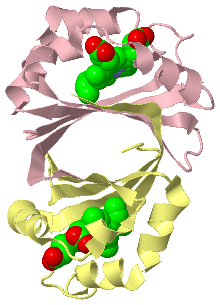
Ligands, Modified Residues, Ions (1, 4)
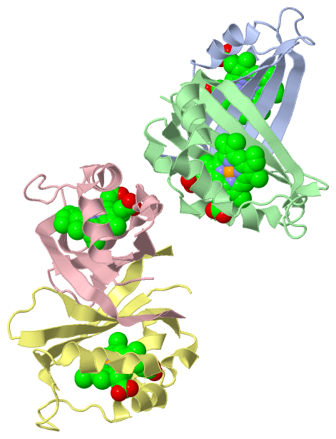
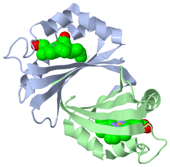
Asymmetric Unit (1, 4)
|
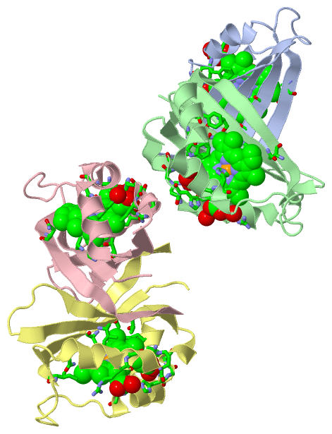
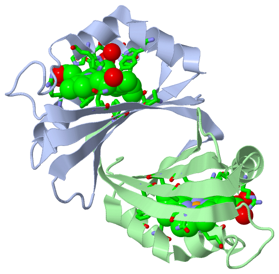
Sites (4, 4)
Asymmetric Unit (4, 4)
|
SS Bonds (0, 0)| (no "SS Bond" information available for 2ZDO) |
Cis Peptide Bonds (0, 0)| (no "Cis Peptide Bond" information available for 2ZDO) |
SAPs(SNPs)/Variants (0, 0)| (no "SAP(SNP)/Variant" information available for 2ZDO) |
PROSITE Motifs (1, 4)
Asymmetric Unit (1, 4)
|
||||||||||||||||||||||||||||||||||||||||||||||||||||||||||||||||||||||||
Exons (0, 0)| (no "Exon" information available for 2ZDO) |
Sequences/Alignments
Asymmetric UnitChain A from PDB Type:PROTEIN Length:108 aligned with HDOX1_STAAN | Q7A649 from UniProtKB/Swiss-Prot Length:107 Alignment length:108 1 | 9 19 29 39 49 59 69 79 89 99 HDOX1_STAAN - -MKFMAENRLTLTKGTAKDIIERFYTRHGIETLEGFDGMFVTQTLEQEDFDEVKILTVWKSKQAFTDWLKSDVFKAAHKHVRSKNEDESSPIINNKVITYDIGYSYMK 107 SCOP domains d2zdoa_ A: automated matches SCOP domains CATH domains 2zdoA00 A:0-107 [code=3.30.70.900, no name defined] CATH domains Pfam domains ------------------------------------------------------------------------------------------------------------ Pfam domains SAPs(SNPs) ------------------------------------------------------------------------------------------------------------ SAPs(SNPs) PROSITE ---ABM PDB: A:3-92 UniProt: 3-92 --------------- PROSITE Transcript ------------------------------------------------------------------------------------------------------------ Transcript 2zdo A 0 TMKFMAEARLTLTKGTAKDIIERFYTRHGIETLEGFDGMFVTQTLEQEDFDEVKILTVWKSKQAFTDWLKSDVFKAAHKHVRSKNEDESSPIINNKVITYDIGYSYMK 107 9 19 29 39 49 59 69 79 89 99 Chain B from PDB Type:PROTEIN Length:108 aligned with HDOX1_STAAN | Q7A649 from UniProtKB/Swiss-Prot Length:107 Alignment length:108 1 | 9 19 29 39 49 59 69 79 89 99 HDOX1_STAAN - -MKFMAENRLTLTKGTAKDIIERFYTRHGIETLEGFDGMFVTQTLEQEDFDEVKILTVWKSKQAFTDWLKSDVFKAAHKHVRSKNEDESSPIINNKVITYDIGYSYMK 107 SCOP domains d2zdob_ B: automated matches SCOP domains CATH domains 2zdoB00 B:0-107 [code=3.30.70.900, no name defined] CATH domains Pfam domains ------------------------------------------------------------------------------------------------------------ Pfam domains SAPs(SNPs) ------------------------------------------------------------------------------------------------------------ SAPs(SNPs) PROSITE ---ABM PDB: B:3-92 UniProt: 3-92 --------------- PROSITE Transcript ------------------------------------------------------------------------------------------------------------ Transcript 2zdo B 0 TMKFMAEARLTLTKGTAKDIIERFYTRHGIETLEGFDGMFVTQTLEQEDFDEVKILTVWKSKQAFTDWLKSDVFKAAHKHVRSKNEDESSPIINNKVITYDIGYSYMK 107 9 19 29 39 49 59 69 79 89 99 Chain C from PDB Type:PROTEIN Length:108 aligned with HDOX1_STAAN | Q7A649 from UniProtKB/Swiss-Prot Length:107 Alignment length:108 1 | 9 19 29 39 49 59 69 79 89 99 HDOX1_STAAN - -MKFMAENRLTLTKGTAKDIIERFYTRHGIETLEGFDGMFVTQTLEQEDFDEVKILTVWKSKQAFTDWLKSDVFKAAHKHVRSKNEDESSPIINNKVITYDIGYSYMK 107 SCOP domains d2zdoc_ C: automated matches SCOP domains CATH domains 2zdoC00 C:0-107 [code=3.30.70.900, no name defined] CATH domains Pfam domains ------------------------------------------------------------------------------------------------------------ Pfam domains SAPs(SNPs) ------------------------------------------------------------------------------------------------------------ SAPs(SNPs) PROSITE ---ABM PDB: C:3-92 UniProt: 3-92 --------------- PROSITE Transcript ------------------------------------------------------------------------------------------------------------ Transcript 2zdo C 0 TMKFMAEARLTLTKGTAKDIIERFYTRHGIETLEGFDGMFVTQTLEQEDFDEVKILTVWKSKQAFTDWLKSDVFKAAHKHVRSKNEDESSPIINNKVITYDIGYSYMK 107 9 19 29 39 49 59 69 79 89 99 Chain D from PDB Type:PROTEIN Length:109 aligned with HDOX1_STAAN | Q7A649 from UniProtKB/Swiss-Prot Length:107 Alignment length:109 1 | 8 18 28 38 48 58 68 78 88 98 HDOX1_STAAN - --MKFMAENRLTLTKGTAKDIIERFYTRHGIETLEGFDGMFVTQTLEQEDFDEVKILTVWKSKQAFTDWLKSDVFKAAHKHVRSKNEDESSPIINNKVITYDIGYSYMK 107 SCOP domains d2zdod_ D: automated matches SCOP domains CATH domains 2zdoD00 D:-1-107 [code=3.30.70.900, no name defined] CATH domains Pfam domains ------------------------------------------------------------------------------------------------------------- Pfam domains SAPs(SNPs) ------------------------------------------------------------------------------------------------------------- SAPs(SNPs) PROSITE ----ABM PDB: D:3-92 UniProt: 3-92 --------------- PROSITE Transcript ------------------------------------------------------------------------------------------------------------- Transcript 2zdo D -1 STMKFMAEARLTLTKGTAKDIIERFYTRHGIETLEGFDGMFVTQTLEQEDFDEVKILTVWKSKQAFTDWLKSDVFKAAHKHVRSKNEDESSPIINNKVITYDIGYSYMK 107 8 18 28 38 48 58 68 78 88 98
|
||||||||||||||||||||
SCOP Domains (1, 4)
Asymmetric Unit
 |
CATH Domains (1, 4)
Asymmetric Unit
|
Pfam Domains (0, 0)| (no "Pfam Domain" information available for 2ZDO) |
Gene Ontology (11, 11)|
Asymmetric Unit(hide GO term definitions) Chain A,B,C,D (HDOX1_STAAN | Q7A649)
|
||||||||||||||||||||||||||||||||||||||||||||||||||||||||||||||||||||||||||||||||||||
Interactive Views
|
||||||||||||||||||||||||||||||||||||||||||||||||||||||||||||||||||||||||||||||||||||||||||||||||||||||||||||||||||||||||||||||||||||||||||||||||||||||||||||||||||
Still Images
|
||||||||||||||||
Databases
|
||||||||||||||||||||||||||||||||||||||||||||||||||||||||||||||||||||||||||||||||||||||||||||||||||||||||||||||||||||||||||||||||||||||||||||||||||||||||||||||||
Analysis Tools
|
|||||||||||||||||||||||||||||||||||||||||||||||||||||||||||||
Entries Sharing at Least One Protein Chain (UniProt ID)
Related Entries Specified in the PDB File
|
|