Show PDB file:   
         Plain Text   HTML   (compressed file size)
QuickSearch:   
by PDB,NDB,UniProt,PROSITE Code or Search Term(s)  
(-)NMR Structure - manually
(-)NMR Structure - model 1
(-)NMR Structure - all models
collapse expand < >
Image NMR Structure - manually
NMR Structure - manually  (Jmol Viewer)
Image NMR Structure - model 1
NMR Structure - model 1  (Jmol Viewer)
Image NMR Structure - all models
NMR Structure - all models  (Jmol Viewer)

(-) Description

Title :  SOLUTION STRUCTURE OF THE THIRD CADHERIN DOMAIN FROM HUMAN PROTOCADHERIN 7
 
Authors :  K. Seimiya, M. Yoshida, F. Hayashi, S. Yokoyama, Riken Structural Genomics/Proteomics Initiative (Rsgi)
Date :  03 Apr 07  (Deposition) - 09 Oct 07  (Release) - 24 Feb 09  (Revision)
Method :  SOLUTION NMR
Resolution :  NOT APPLICABLE
Chains :  NMR Structure  :  A  (20x)
Keywords :  Brain-Heart Protocadherin, Bh-Pcdh, Beta-Sandwich, Structural Genomics, Nppsfa, National Project On Protein Structural And Functional Analyses, Riken Structural Genomics/Proteomics Initiative, Rsgi, Cell Adhesion (Keyword Search: [Gene Ontology, PubMed, Web (Google))
 
Reference :  K. Seimiya, M. Yoshida, F. Hayashi, S. Yokoyama
Solution Structure Of The Third Cadherin Domain From Human Protocadherin 7
To Be Published
PubMed: search
(for further references see the PDB file header)

(-) Compounds

Molecule 1 - PROTOCADHERIN-7
    ChainsA
    EngineeredYES
    Expression System PlasmidP060515-20
    Expression System Vector TypePLASMID
    FragmentCADHERIN
    GenePCDH7, BHPCDH
    Organism CommonHUMAN
    Organism ScientificHOMO SAPIENS
    Organism Taxid9606
    Other DetailsCELL FREE PROTEIN SYNTHESIS
    SynonymBRAIN-HEART PROTOCADHERIN, BH-PCDH

 Structural Features

(-) Chains, Units

  
NMR Structure (20x)

Summary Information (see also Sequences/Alignments below)

(-) Ligands, Modified Residues, Ions  (0, 0)

(no "Ligand,Modified Residues,Ions" information available for 2YST)

(-) Sites  (0, 0)

(no "Site" information available for 2YST)

(-) SS Bonds  (0, 0)

(no "SS Bond" information available for 2YST)

(-) Cis Peptide Bonds  (1, 20)
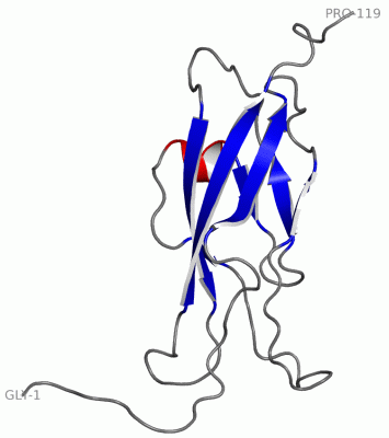
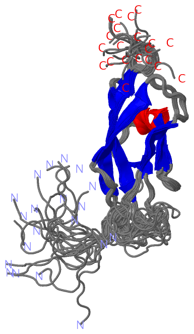
NMR Structure
No.ModelResidues
11, 2, 3, 4, 5, 6, 7, 8, 9, 10, 11, 12, 13, 14, 15, 16, 17, 18, 19, 20Gln A:98 -Pro A:99

 Sequence-Structure Mapping

(-) SAPs(SNPs)/Variants  (0, 0)

(no "SAP(SNP)/Variant" information available for 2YST)

(-) PROSITE Motifs  (2, 3)

NMR Structure (2, 3)
 PROSITEUniProtKBPDB
No.IDACDescriptionIDLocationCountLocation
1CADHERIN_2PS50268 Cadherins domain profile.PCDH7_HUMAN52-143
144-308
309-415
432-535
536-639
640-742
745-862
  1-
-
A:15-119
-
-
-
-
2CADHERIN_1PS00232 Cadherin domain signature.PCDH7_HUMAN131-141
296-306
403-413
523-533
627-637
730-740
  2-
A:8-12
A:109-119
-
-
-

(-) Exons   (1, 1)

NMR Structure (1, 1)
 ENSEMBLUniProtKBPDB
No.Transcript IDExonExon IDGenome LocationLengthIDLocationLengthCountLocationLength
1.1aENST000003617621aENSE00001435176chr4:30722037-307262184182PCDH7_HUMAN1-105810581A:1-119 (gaps)204
1.2ENST000003617622ENSE00001435501chr4:30732375-307335901216PCDH7_HUMAN1059-1069110--

(-) Sequences/Alignments

NMR Structure
   Reformat: Number of residues per line =  ('0' or empty: single-line sequence representation)
  Number of residues per labelling interval =   
  UniProt sequence: complete  aligned part    
   Show mapping: SCOP domains CATH domains Pfam domains Secondary structure (by author)
SAPs(SNPs) PROSITE motifs Exons
(details for a mapped element are shown in a popup box when the mouse pointer rests over it)
Chain A from PDB  Type:PROTEIN  Length:119
 aligned with PCDH7_HUMAN | O60245 from UniProtKB/Swiss-Prot  Length:1069

    Alignment length:204
                                   219       229       239       249       259       269       279       289       299       309       319       329       339       349       359       369       379       389       399       409    
          PCDH7_HUMAN   210 GGNGASGGGSGGSKRRLDASEGGGGTNPGGRSSVFELQVADTPDGEKQPQLIVKGALDREQRDSYELTLRVRDGGDPPRSSQAILRVLITDVNDNSPRFEKSVYEADLAENSAPGTPILQLRAADLDVGVNGQIEYVFGAATESVRRLLRLDETSGWLSVLHRIDREEVNQLRFTVMARDRGQPPKTDKATVVLNIKDENDNVP 413
               SCOP domains ------------------------------------------------------------------------------------------------------------------------------------------------------------------------------------------------------------ SCOP domains
               CATH domains ------------------------------------------------------------------------------------------------------------------------------------------------------------------------------------------------------------ CATH domains
               Pfam domains ------------------------------------------------------------------------------------------------------------------------------------------------------------------------------------------------------------ Pfam domains
         Sec.struct. author .......-------------------------------------------------------------------------------------..........eeeeeee.......eeee................ee.....hhhhheee.....eeee..........eeeeeeee.........eeeeeeeeee....... Sec.struct. author
                 SAPs(SNPs) ------------------------------------------------------------------------------------------------------------------------------------------------------------------------------------------------------------ SAPs(SNPs)
                PROSITE (1) CADHERIN_2  PDB: - UniProt: 144-308                                                                CADHERIN_2  PDB: A:15-119 UniProt: 309-415                                                                PROSITE (1)
                PROSITE (2) --------------------------------------------------------------------------------------CADHERIN_1 ------------------------------------------------------------------------------------------------CADHERIN_1  PROSITE (2)
               Transcript 1 Exon 1.1a  PDB: A:1-119 (gaps) UniProt: 1-1058 [INCOMPLETE]                                                                                                                                                  Transcript 1
                 2yst A   1 GSSGSSG-------------------------------------------------------------------------------------NDNSPRFEKSVYEADLAENSAPGTPILQLRAADLDVGVNGQIEYVFGAATESVRRLLRLDETSGWLSVLHRIDREEVNQLRFTVMARDRGQPPKTDKATVVLNIKDENDNVP 119
                                  |  -         -         -         -         -         -         -         -         -  |     15        25        35        45        55        65        75        85        95       105       115    
                                  7                                                                                     8                                                                                                               

   Legend:   → Mismatch (orange background)
  - → Gap (green background, '-', border residues have a numbering label)
    → Modified Residue (blue background, lower-case, 'x' indicates undefined single-letter code, labelled with number + name)
  x → Chemical Group (purple background, 'x', labelled with number + name, e.g. ACE or NH2)
  extra numbering lines below/above indicate numbering irregularities and modified residue names etc., number ends below/above '|'

 Classification and Annotation

(-) SCOP Domains  (0, 0)

(no "SCOP Domain" information available for 2YST)

(-) CATH Domains  (0, 0)

(no "CATH Domain" information available for 2YST)

(-) Pfam Domains  (0, 0)

(no "Pfam Domain" information available for 2YST)

(-) Gene Ontology  (9, 9)

NMR Structure(hide GO term definitions)
Chain A   (PCDH7_HUMAN | O60245)
molecular function
    GO:0005509    calcium ion binding    Interacting selectively and non-covalently with calcium ions (Ca2+).
biological process
    GO:0007155    cell adhesion    The attachment of a cell, either to another cell or to an underlying substrate such as the extracellular matrix, via cell adhesion molecules.
    GO:0007156    homophilic cell adhesion via plasma membrane adhesion molecules    The attachment of a plasma membrane adhesion molecule in one cell to an identical molecule in an adjacent cell.
    GO:0002576    platelet degranulation    The regulated exocytosis of secretory granules containing preformed mediators such as histamine and serotonin by a platelet.
cellular component
    GO:0016021    integral component of membrane    The component of a membrane consisting of the gene products and protein complexes having at least some part of their peptide sequence embedded in the hydrophobic region of the membrane.
    GO:0005887    integral component of plasma membrane    The component of the plasma membrane consisting of the gene products and protein complexes having at least some part of their peptide sequence embedded in the hydrophobic region of the membrane.
    GO:0016020    membrane    A lipid bilayer along with all the proteins and protein complexes embedded in it an attached to it.
    GO:0005886    plasma membrane    The membrane surrounding a cell that separates the cell from its external environment. It consists of a phospholipid bilayer and associated proteins.
    GO:0031092    platelet alpha granule membrane    The lipid bilayer surrounding the platelet alpha granule.

 Visualization

(-) Interactive Views

NMR Structure
  Complete Structure
    Jena3D(integrated viewing of ligand, site, SAP, PROSITE, SCOP information)
    WebMol | AstexViewer[tm]@PDBe
(Java Applets, require no local installation except for Java; loading may be slow)
    STRAP
(Java WebStart application, automatic local installation, requires Java; full application with system access!)
    RasMol
(require local installation)
    Molscript (VRML)
(requires installation of a VRML viewer; select preferred view via VRML and generate a mono or stereo PDF format file)
 
  Ligands, Modified Residues, Ions
(no "Ligands, Modified Residues, Ions" information available for 2yst)
 
  Sites
(no "Sites" information available for 2yst)
 
  Cis Peptide Bonds
    Gln A:98 - Pro A:99   [ RasMol ]  
 

(-) Still Images

Jmol
  protein: cartoon or spacefill or dots and stick; nucleic acid: cartoon and stick; ligands: spacefill; active site: stick
Molscript
  protein, nucleic acid: cartoon; ligands: spacefill; active site: ball and stick

 Databases and Analysis Tools

(-) Databases

Access by PDB/NDB ID
  2yst
    Family and Domain InformationProDom | SYSTERS
    General Structural InformationGlycoscienceDB | MMDB | NDB | OCA | PDB | PDBe | PDBj | PDBsum | PDBWiki | PQS | PROTEOPEDIA
    Orientation in MembranesOPM
    Protein SurfaceSURFACE
    Secondary StructureDSSP (structure derived) | HSSP (homology derived)
    Structural GenomicsGeneCensus
    Structural NeighboursCE | VAST
    Structure ClassificationCATH | Dali | SCOP
    Validation and Original DataBMRB Data View | BMRB Restraints Grid | EDS | PROCHECK | RECOORD | WHAT_CHECK
 
Access by UniProt ID/Accession number
  PCDH7_HUMAN | O60245
    Comparative Protein Structure ModelsModBase
    Genomic InformationEnsembl
    Protein-protein InteractionDIP
    Sequence, Family and Domain InformationInterPro | Pfam | SMART | UniProtKB/SwissProt
 
Access by Enzyme Classificator   (EC Number)
  (no 'Enzyme Classificator' available)
    General Enzyme InformationBRENDA | EC-PDB | Enzyme | IntEnz
    PathwayKEGG | MetaCyc
 
Access by Disease Identifier   (MIM ID)
  (no 'MIM ID' available)
    Disease InformationOMIM
 
Access by GenAge ID
  (no 'GenAge ID' available)
    Age Related InformationGenAge

(-) Analysis Tools

Access by PDB/NDB ID
    Domain InformationXDom
    Interatomic Contacts of Structural UnitsCSU
    Ligand-protein ContactsLPC
    Protein CavitiescastP
    Sequence and Secondary StructurePDBCartoon
    Structure AlignmentSTRAP(Java WebStart application, automatic local installation, requires Java; full application with system access!)
    Structure and Sequence BrowserSTING
 
Access by UniProt ID/Accession number
  PCDH7_HUMAN | O60245
    Protein Disorder PredictionDisEMBL | FoldIndex | GLOBPLOT (for more information see DisProt)

 Related Entries

(-) Entries Sharing at Least One Protein Chain (UniProt ID)

(no "Entries Sharing at Least One Protein Chain" available for 2YST)

(-) Related Entries Specified in the PDB File

(no "Related Entries Specified in the PDB File" available for 2YST)